Ophthalmic Imaging
In the recent decade, ophthalmic imaging has proven to be a steadily growing field of research. A milestone in ophthalmology was the transfer of Optical Coherence Tomography (OCT) from research to clinical use. Compared to conventional fundus photography, OCT allows the three dimensional, depth resolved visualization of the human retina, while preserving the non-invasiveness. Recently, a radical change occured in the field of OCT research with the clinical introduction of OCT angiography (OCTA), which further adds dye-free imaging of the underlying vasculature.

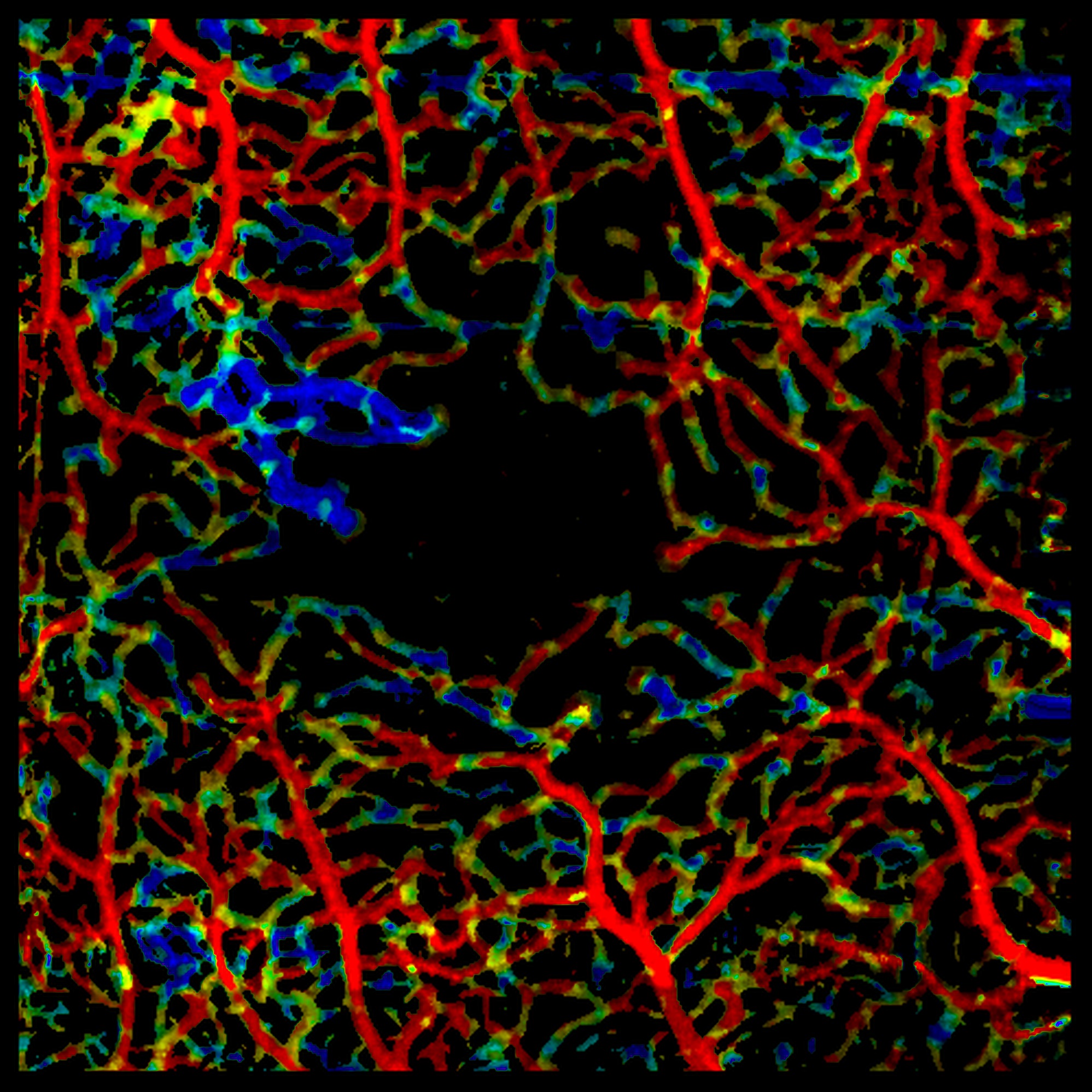
Variable Interscan Time Analysis revealing slower blood flow in neovascularization in a PDR patient.
Research Focus
To facilitate an efficient analysis of this vastly increased amount of information, new processing algorithms are required to support the treating clinician. Our research focus is twofold: On the one hand, we develop advanced motion compensation, shadow artifact compensation and signal reconstruction algorithms to achieve artifact-free OCT(A) signals of best possible quality. On the other hand, we aid accurate image analysis by improving layer and vessel segmentations, categorizing vessels into arteries/veins or pathology. By combining both efforts, we want to improve the understanding of the most prevalent eye diseases, allowing for more accurate treatment and thus improved patient outcome.

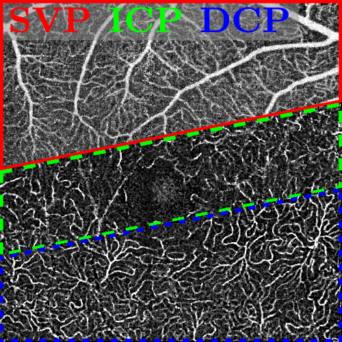
Advanced shadow artifact removal reveals the unique structures of the retinal vascular networks.
Multidisciplinary Collaborations
To enable research with state-of-the-art technology while preserving a close link to the clinical needs, the work of our group is embedded in a multidisciplinary environment including optical engineers at the Massachusetts Institute of Technology, Cambridge, USA and clinicians at the New England Eye Center, Boston, USA and the Department of Ophthalmology at the University Clinic Erlangen.
Participating Scientists

Lennart Husvogt, M. Sc.
- Phone number: +49 9131 85-25247
- Email: lennart.husvogt@fau.de
- Website: https://lme.tf.fau.de/person/husvogt/

Stefan Ploner, M. Sc.
- Phone number: +49 9131 85-28982
- Email: stefan.ploner@fau.de
- Website: https://lme.tf.fau.de/person/ploner/
Projects
4D-OCTA: Temporally resolved 3-D retinal blood flow quantification using advanced motion correction and signal reconstruction in optical coherence tomography angiography
Die optische Kohärenztomographie (OCT) erzeugt volumetrische 3-D-Bilder von Gewebe mit Mikrometerauflösung, indem sie einen Laserstrahl zum Scannen verwendet und die Amplitude und Zeitverzögerung von zurückgestreutem Licht misst. Die OCT hat einen großen Einfluss auf die Augenheilkunde und wurde zu einer Standard-Bildgebungsmodalität für die Diagnose, die Überwachung des Krankheitsverlaufs und das Ansprechen auf die Behandlung sowie für die Untersuchung der Pathogenese von Krankheiten wie diab…
Joint Reco & MoCo for OCT(A): Joint Iterative Reconstruction and Motion Compensation for Optical Coherence Tomography Angiography
Publications
- Lee B., Moult EM., Ploner S., Alibhai AY., Rebhun CB., Moreira-Neto C., Husvogt L., Maier A., Wollstein G., Schuman J., Waheed NK., Duker JS., Fujimoto JG.:
Parallel Variable Interscan Time Analysis (VISTA) OCTA and en Face Doppler OCT of Optic Disc and Peripapillary Vasculature
ARVO Annual Meeting 2017 (Baltimore, MD, USA, May 7, 2017 - May 11, 2017)
In: Investigative Ophthalmology & Visual Science 2017
URL: https://www5.informatik.uni-erlangen.de/Forschung/Publikationen/2017/Lee17-PVI.pdf
BibTeX: Download - Moult EM., Ploner S., Rebhun CB., Alibhai AY., Lee B., Moreira-Neto C., Novais EA., Schottenhamml J., Husvogt L., Maier A., Rosenfeld P., Duker JS., Waheed NK., Fujimoto JG.:
Variable Interscan Time Analysis (VISTA-) Optical Coherence Tomography Angiography (OCTA) of Blood Flow Speeds in Eyes with Diabetic Retinopathy
ARVO Annual Meeting 2017 (Baltimore, MD, USA, May 7, 2017 - May 11, 2017)
In: Investigative Ophthalmology & Visual Science, ARVO: 2017
URL: https://iovs.arvojournals.org/article.aspx?articleid=2637998&resultClick=1
BibTeX: Download - Giacomelli M., Yoshitake T., Husvogt L., Cahill L., Ahsen O., Vardeh H., Sheykin Y., Faulkner-Jones BE., Hornegger J., Brooker J., Cable AE., Connolly JL., Fujimoto JG.:
Design of a portable wide field of view GPU-accelerated multiphoton imaging system for real-time imaging of breast surgical specimens
SPIE Photonics West (San-Francisco, CA, February 14, 2016 - February 16, 2016)
In: Multiphoton Microscopy in the Biomedical Sciences XVI 2016
DOI: 10.1117/12.2209241
URL: https://www.spiedigitallibrary.org/conference-proceedings-of-spie/9712/1/Design-of-a-portable-wide-field-of-view-GPU-accelerated/10.1117/12.2209241.full?SSO=1
BibTeX: Download
