

Global Breast Cancer Burden and the Clinical Care Pathway
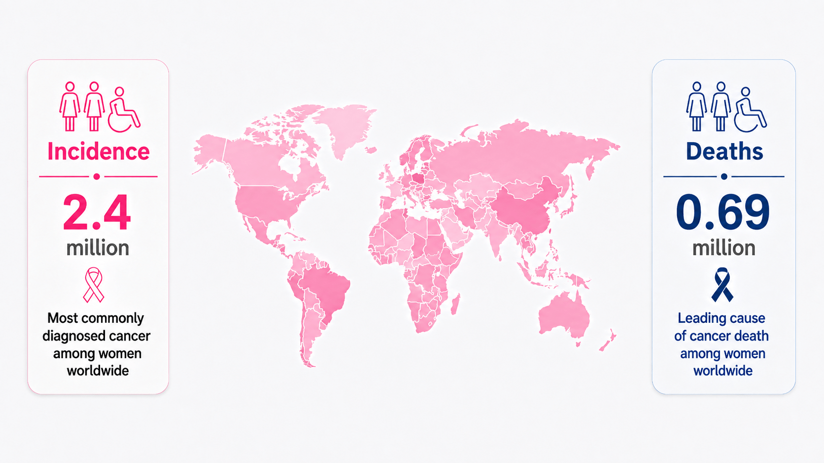
Every year, approximately 2.4 million new breast cancer cases and 0.69 million breast cancer deaths occur worldwide.
The scale of this global burden highlights the importance of an effective clinical pathway spanning early detection, diagnostic imaging, pathological assessment, staging and personalized treatment.
Annual Global Breast Cancer Burden
From Screening to Personalized Treatment
Breast care follows a connected pathway in which screening findings, mammography, additional imaging, tissue biopsy and pathological analysis contribute to clinical decision-making and treatment planning.
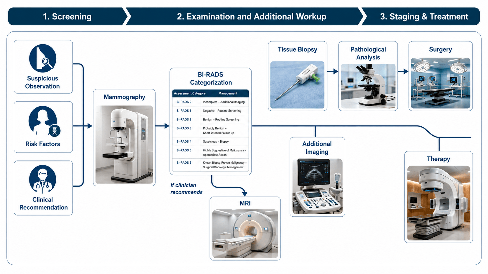
The incidence and mortality values shown are approximate annual global figures. Individual care pathways may vary according to imaging findings, pathology, tumour biology, disease stage and clinical circumstances. Diagnostic and treatment decisions are made by the responsible multidisciplinary clinical team.
Breast Imaging AI in Action
Explore three research demonstrators developed at the Pattern Recognition Lab for AI-assisted breast-image analysis.
The showcase presents MammoLocate for mammographic lesion localization, MammoGPT for report generation and visual question answering, and BE-WISE for explainable breast MRI analysis.
These demonstrators represent ongoing research and are intended to illustrate experimental AI-assisted breast-imaging methods rather than systems for independent clinical decision-making.
“If current rates continue, by 2050 there will be 3.2 million new breast cancer cases and 1.1 million breast cancer-related deaths per year.”
International Agency for Research on Cancer, 2025
Research Across the Breast-Care Continuum
Our breast-related research spans the complete imaging pathway—from acquisition and reconstruction to clinical image analysis, computational pathology and multimodal artificial intelligence.
Explore the research areas below and open each section to view the corresponding publications.
Mammography AI & Screening
Detection, classification, density assessment and clinically oriented mammography analysis.
PRL develops learning-based methods for mammographic density assessment, calcification analysis, abnormality classification and lesion detection. The research covers conventional, contrast-enhanced and multiview mammography, together with AI-assisted screening studies. A central objective is to improve robustness across datasets, breast densities and clinical populations while addressing clinically relevant operating conditions such as high specificity.
View publications
- Bhandary Panambur, A., Bhat, S., Yu, H., Madhu, P., Bayer, S., & Maier, A. (2025). Attention-guided erasing for enhanced transfer learning in breast abnormality classification. International Journal of Computer Assisted Radiology and Surgery. https://doi.org/10.1007/s11548-024-03317-6
BibTeX: Download
- Bhandary Panambur, A. (2024). Enhancing mammography screening sensitivity with AI-assistance: Evidence from a Vietnamese study cohort. ESR Eurosafe Imaging.
BibTeX: Download
- Bhandary Panambur, A., Madhu, P., Bayer, S., & Maier, A. (2024). Enhancing downstream classification of breast abnormalities in contrast enhanced spectral mammography using a neighborhood representation loss. In Weijie Chen, Susan M. Astley (Eds.), Medical Imaging 2024: Computer-Aided Diagnosis. San Diego, US: SPIE.
BibTeX: Download
- Bhat, S., Mansoor, A., Georgescu, B., Bhandary Panambur, A., Ghesu, F.C., Islam, S.,... Maier, A. (2023). AUCReshaping: improved sensitivity at high-specificity. Scientific Reports, 13. https://doi.org/10.1038/s41598-023-48482-x
BibTeX: Download
- El-Ghoussani, A., Rodriguez Salas, D., Seuret, M., & Maier, A. (2022). GAN-based Augmentation of Mammograms to Improve Breast Lesion Detection. In Klaus Maier-Hein, Thomas M. Deserno, Heinz Handels, Andreas Maier, Christoph Palm, Thomas Tolxdorff (Eds.), Informatik aktuell (pp. 321-326). Heidelberg, DEU, DE: Wiesbaden: Springer Science and Business Media Deutschland GmbH.
BibTeX: Download
- Bhandary Panambur, A., Madhu, P., & Maier, A. (2022). Effect of Random Histogram Equalization on Breast Calcification Analysis Using Deep Learning. In Proceedings of the German Workshop on Medical Image Computing, 2022 (pp. 173-178). Springer Science and Business Media Deutschland GmbH.
BibTeX: Download
- Kaiser, N., Fieselmann, A., Vesal, S., Ravikumar, N., Ritschl, L., Kappler, S., & Maier, A. (2019). Mammographic breast density classification using a deep neural network: assessment based on inter-observer variability. In MEDICAL IMAGING 2019: IMAGE PERCEPTION, OBSERVER PERFORMANCE, AND TECHNOLOGY ASSESSMENT. San Diego, CA, US: BELLINGHAM: SPIE-INT SOC OPTICAL ENGINEERING.
BibTeX: Download
- Schebesch, F., Unberath, M., Andersen, I., & Maier, A. (2017). Breast density assessment using wavelet features on mammograms. In Thomas M. Deserno, Heinz Handels, Thomas Tolxdorff, Hans-Peter Meinzer (Eds.), Informatik aktuell (pp. 38-43). Berlin, DEU: Kluwer Academic Publishers.
BibTeX: Download
Acquisition, Reconstruction & Image Quality
From imaging physics and acquisition to reliable image formation.
Reliable image analysis begins with reliable image formation. This research addresses mammographic compression, denoising, digital breast tomosynthesis resolution, physical phantoms and virtual clinical trials. By combining imaging physics with computational reconstruction and learning-based methods, PRL investigates how acquisition parameters influence image quality, anatomical detail and downstream analysis.
View publications
- Hertel, M., Liu, C., Song, H., Golatta, M., Kappler, S., Nanke, R.,... Rose, G. (2023). Clinical prototype implementation enabling an improved day-to-day mammography compression. Physica Medica, 106. https://doi.org/10.1016/j.ejmp.2023.102524
BibTeX: Download
- Eckert, D., Ritschl, L., Herbst, M., Wicklein, J., Vesal, S., Kappler, S.,... Stober, S. (2022). Deep learning based denoising of mammographic x-ray images: an investigation of loss functions and their detail-preserving properties. In Wei Zhao, Lifeng Yu (Eds.), Progress in Biomedical Optics and Imaging - Proceedings of SPIE. Virtual, Online: SPIE.
BibTeX: Download
- Eckert, D., Vesal, S., Ritschl, L., Kappler, S., & Maier, A. (2020). Deep learning-based denoising of mammographic images using physics-driven data augmentation. In Thomas Tolxdorff, Thomas M. Deserno, Heinz Handels, Andreas Maier, Klaus H. Maier-Hein, Christoph Palm (Eds.), Informatik aktuell (pp. 94-100). Berlin, DE: Springer.
BibTeX: Download
- Luckner, C., Schebesch, F., Mertelmeier, T., Fieselmann, A., Maier, A., & Ritschl, L. (2018). Towards an analytic model: Describing the effect of scan angle and slice thickness on the in-plane spatial resolution of calcications in digital breast tomosynthesis. In Proc. of SPIE (pp. 107181R). Atlanta, GA, USA.
BibTeX: Download
- Luckner, C., Schebesch, F., Syben-Leisner, C., Mertelmeier, T., Maier, A., & Ritschl, L. (2018). On the Influence of Acquisition Angle and Slice Thickness on the in-plane Spatial Resolution of Calcifications in Digital Breast Tomosynthesis. In Proceedings of the Fifth International Conference on Image Formation in X-Ray Computed Tomography (pp. 147-150). Salt Lake City, USA.
BibTeX: Download
- Schebesch, F., Magdalena, H., Mertelmeier, T., Maier, A., & Ritschl, L. (2018). A Hybrid Approach for Virtual Clinical Trials for Mammographic Imaging. In Proc. of SPIE (pp. 107180Z). Atlanta, GA, USA: SPIE.
BibTeX: Download
- Hanif, S., Schebesch, F., Jerebko, A., Ritschl, L., Mertelmeier, T., & Maier, A. (2017). Lesion Ground Truth Estimation for a Physical Breast Phantom. In Bildverarbeitung für die Medizin 2017 - Algorithmen, Systeme, Anwendungen (pp. 243-248). Heidelberg: Kluwer Academic Publishers.
BibTeX: Download
Breast MRI
Lesion analysis, image quality, synthetic imaging and explainable AI.
Breast MRI research at PRL covers lesion segmentation, artifact detection, image-quality assessment, background parenchymal enhancement and clinical decision support. More recent work investigates virtual contrast enhancement, generative imaging and weakly supervised explanations. The programme combines medical image analysis and deep learning to support more reliable acquisition, interpretation and communication of MRI findings. Much of this work is conducted in close collaboration with the tMRI Lab.
For more information, visit: https://www.mr-physik.med.fau.de/lab/lab-transformative-oncologic-imaging-troi/
View publications
- Bhandary Panambur, A., Nguyen, T.-T., Bayer, S., & Maier, A. (2026). Breast MRI Evaluation with Weakly-informed Slice-level Explanation. In Proceedings of the Bildverarbeitung für die Medizin 2026 (pp. 10-17). Lübeck, DE.
BibTeX: Download
- George, A., Schreiter, H., Hoßbach, J., Nguyen, T.-T., Horishnyi, I., Ehring, C.,... Liebert, A. (2025). U-Net and GAN for Virtual Contrast in Breast MRI: How Do They Compare to Real Contrast Images? In Christoph Palm, Katharina Breininger, Thomas Deserno, Heinz Handels, Andreas Maier, Klaus H. Maier-Hein, Thomas M. Tolxdorff (Eds.), Bildverarbeitung für die Medizin 2025. Proceedings, German Conference on Medical Image Computing, Regensburg March 09-11, 2025 (pp. 277-282). Regensburg, DE: Cham: Springer.
BibTeX: Download
- Okolie, A., Dirrichs, T., Huck, L., Nebelung, S., Tayebi Arasteh, S., Nolte, T.,... Truhn, D. (2024). Accelerating breast MRI acquisition with generative AI models. European Radiology. https://doi.org/10.1007/s00330-024-10853-x
BibTeX: Download
- Kapsner, L., Folle, L., Hadler, D., Eberle, J., Balbach, E., Liebert, A.,... Bickelhaupt, S. (2024). Lesion-conditioning of synthetic MRI-derived subtraction-MIPs of the breast using a latent diffusion model. Scientific Reports, 14(1). https://doi.org/10.1038/s41598-024-56853-1
BibTeX: Download
- Müller-Franzes, G., Huck, L., Tayebi Arasteh, S., Khader, F., Han, T., Schulz, V.,... Truhn, D. (2023). Using machine learning to reduce the need for contrast agents in breast MRI through synthetic images. Radiology, 307(3). https://doi.org/10.1148/radiol.222211
BibTeX: Download
- Das, B., Kapsner, L., Ohlmeyer, S., Laun, F.B., Maier, A., Uder, M.,... Liebert, A. (2023). Detection and prediction of background parenchymal enhancement on breast MRI using deep learning. In Proceedings of the Joint Annual Meeting ISMRM-ESMRMB & ISMRT 31st Annual Meeting. London, GB.
BibTeX: Download
- Kapsner, L., Balbach, E., Folle, L., Laun, F.B., Nagel, A.M., Liebert, A.,... Bickelhaupt, S. (2023). Image quality assessment using deep learning in high b-value diffusion-weighted breast MRI. Scientific Reports, 13(1). https://doi.org/10.1038/s41598-023-37342-3
BibTeX: Download
- Kapsner, L., Ohlmeyer, S., Folle, L., Laun, F.B., Nagel, A.M., Liebert, A.,... Bickelhaupt, S. (2022). Automated artifact detection in abbreviated dynamic contrast-enhanced (DCE) MRI-derived maximum intensity projections (MIPs) of the breast. European Radiology. https://doi.org/10.1007/s00330-022-08626-5
BibTeX: Download
- Ellmann, S., Wenkel, E., Dietzel, M., Bielowski, C., Vesal, S., Maier, A.,... Bäuerle, T. (2020). Implementation of machine learning into clinical breast MRI: Potential for objective and accurate decision-making in suspicious breast masses. PLoS ONE, 1-15, 1-15. https://doi.org/10.1371/journal.pone.0228446
BibTeX: Download
- Vesal, S., Ravikumar, N., Ellmann, S., & Maier, A. (2018). Comparative analysis of unsupervised algorithms for breast MRI lesion segmentation. In Heinz Handels, Thomas Tolxdorff, Thomas M. Deserno, Klaus H. Maier-Hein, Andreas Maier, Christoph Palm (Eds.), Informatik aktuell (pp. 257-262). Erlangen, DEU: Springer Berlin Heidelberg.
BibTeX: Download
- Vesal, S., Diaz-Pinto, A., Ravikumar, N., Ellmann, S., Davari, A., & Maier, A. (2017). Semi-Automatic Algorithm for Breast MRI Lesion Segmentation Using Marker-Controlled Watershed Transformation. In 2017 IEEE Nuclear Science Symposium and Medical Imaging Conference Record (NSS/MIC) (pp. tbd). Atlanta, Georgia, USA.
BibTeX: Download
Ultrasound & Hybrid Imaging
Automated ultrasound, reconstruction and complementary multimodal imaging.
PRL explores automated breast-ultrasound analysis, speed-of-sound reconstruction and weakly supervised lesion detection. Hybrid ultrasound–X-ray concepts investigate how complementary physical measurements can be integrated into mammographic imaging systems. This research aims to improve tissue characterization and extract information that may not be visible through a single imaging modality alone.
View publications
- Rodriguez Salas, D., Öttl, M., Seuret, M., Packhäuser, K., & Maier, A. (2023). Using Forestnets for Partial Fine-Tuning Prior to Breast Cancer Detection in Ultrasounds. In 2023 IEEE 20th International Symposium on Biomedical Imaging (ISBI). Cartagena, Colombia: IEEE Computer Society.
BibTeX: Download
- Rodriguez Salas, D., Seuret, M., Vesal, S., & Maier, A. (2021). Ultrasound Breast Lesion Detection using Extracted Attention Maps from a Weakly Supervised Convolutional Neural Network. In Palm, Christoph; Deserno, Thomas M.; Handels, Heinz;Maier, Andreas; Maier-Hein, Klaus;Tolxdorff, Thomas. (Eds.), Bildverarbeitung für die Medizin 2021 (pp. 282-287). Regensburg, DE: Wiesbaden: Springer Vieweg.
BibTeX: Download
- Khun Jush, F., Biele, M., Dueppenbecker, P.M., Schmidt, O., & Maier, A. (2020). DNN-based Speed-of-Sound Reconstruction for Automated Breast Ultrasound. In 2020 IEEE International Ultrasonics Symposium (IUS) (pp. 1-7). Las Vegas, NV, US: IEEE.
BibTeX: Download
- Li, Q., Luckner, C., Hertel, M., Radicke, M., & Maier, A. (2019). Combining Ultrasound and X-Ray Imaging for Mammography: A Prototype Design. In Thomas M. Deserno, Andreas Maier, Christoph Palm, Heinz Handels, Klaus H. Maier-Hein, Thomas Tolxdorff (Eds.), Informatik aktuell (pp. 245-250). Lübeck, DE: Springer Berlin Heidelberg.
BibTeX: Download
- Li, Q., Luckner, C., Hertel, M., Radicke, M., & Maier, A. (2019). Combining Ultrasound and X-Ray Imaging for Mammography. In Bildverarbeitung für die Medizin 2019. (pp. 245--250). Springer.
BibTeX: Download
Histopathology, Microscopy & Biomarkers
From tissue morphology to computational biomarkers and cancer prognosis.
Breast cancer research at PRL extends from radiological imaging to tissue-level evidence. The portfolio includes histology classification, automated mitosis detection, whole-slide imaging datasets, multiphoton microscopy and high-dimensional immune-cell analysis. These studies connect computational morphology and biological biomarkers with tumour characterization, prognosis and translational cancer research.
View publications
- Stathonikos, N., Aubreville, M., de Vries, S., Wilm, F., Bertram, C.A., Veta, M., & van Diest, P.J. (2024). Breast cancer survival prediction using an automated mitosis detection pipeline. Journal of Pathology: Clinical Research, 10(6). https://doi.org/10.1002/2056-4538.70008
BibTeX: Download
- Heger, L., Heidkamp, G.F., Amon, L., Nimmerjahn, F., Bäuerle, T., Maier, A.,... Dudziak, D. (2024). Unbiased high-dimensional flow cytometry identified NK and DC immune cell signature in Luminal A-type and triple negative breast cancer. OncoImmunology, 13(1). https://doi.org/10.1080/2162402X.2023.2296713
BibTeX: Download
- Aubreville, M., Bertram, C.A., Donovan, T.A., Marzahl, C., Maier, A., & Klopfleisch, R. (2021). A Completely Annotated Whole Slide Image Dataset of Canine Breast Cancer to Aid Human Breast Cancer Research. In Christoph Palm, Heinz Handels, Klaus Maier-Hein, Thomas M. Deserno, Andreas Maier, Thomas Tolxdorff (Eds.), Informatik aktuell (pp. 198-). Regensburg, DE: Springer Science and Business Media Deutschland GmbH.
BibTeX: Download
- Aubreville, M., Bertram, C.A., Donovan, T.A., Marzahl, C., Maier, A., & Klopfleisch, R. (2020). A completely annotated whole slide image dataset of canine breast cancer to aid human breast cancer research. Scientific Data, 7(1). https://doi.org/10.1038/s41597-020-00756-z
BibTeX: Download
- Vesal, S., Ravikumar, N., Davari, A., Ellmann, S., & Maier, A. (2018). Classification of Breast Cancer Histology Images Using Transfer Learning. In ICIAR 2018: Image Analysis and Recognition (pp. 812--819). Springer.
BibTeX: Download
- Giacomelli, M., Yoshitake, T., Husvogt, L., Cahill, L., Ahsen, O., Vardeh, H.,... Fujimoto, J.G. (2016). Design of a portable wide field of view GPU-accelerated multiphoton imaging system for real-time imaging of breast surgical specimens. In Multiphoton Microscopy in the Biomedical Sciences XVI (pp. 0-0). San-Francisco, CA, US: SPIE.
BibTeX: Download
Risk Assessment & Image-Guided Therapy
Risk stratification, CT analysis, treatment planning and adaptive intervention.
Beyond primary breast imaging, PRL investigates opportunistic cancer-risk stratification from chest CT, radiation-aware acquisition and adaptive breast brachytherapy. This work combines multiple-instance learning, dose modulation, geometric modelling and image-guided intervention. It broadens the research programme from diagnosis toward prevention, treatment planning and longitudinal patient care.
View publications
- Ni, Y., Panambur, A.B., Liu, C., Nguyen, T.-T., Bayer, S., Juan, H.,... Maier, A. (2026). Opportunistic Breast Cancer Risk Stratification From Low-dose Chest CT Using Multiple Instance Learning. In Bildverarbeitung für die Medizin 2026 (pp. 427-434). Lübeck, DE.
BibTeX: Download
- Dürrbeck, C., Schulz, M., Pflaum, L., Kallis, K., Geimer, T., Abu-Hossin, N.,... Bert, C. (2023). Estimating follow-up CTs from geometric deformations of catheter implants in interstitial breast brachytherapy: A feasibility study using electromagnetic tracking. Medical Physics. https://doi.org/10.1002/mp.16659
BibTeX: Download
- Klein, L., Enzmann, L., Byl, A., Liu, C., Sawall, S., Maier, A.,... Kachelrieß, M. (2022). Organ-Specific vs. Patient Risk-Specific Tube Current Modulation in Thorax CT Scans Covering the Female Breast. In Joseph Webster Stayman (Eds.), Proceedings of SPIE - The International Society for Optical Engineering. Virtual, Online: SPIE.
BibTeX: Download
- Dürrbeck, C., Pflaum, L., Schulz, M., Kallis, K., Geimer, T., Abu-Hossin, N.,... Bert, C. (2021). Implant-based CT estimation towards adaptive breast brachytherapy. In RADIOTHERAPY AND ONCOLOGY (pp. S76-S77). CLARE: ELSEVIER IRELAND LTD.
BibTeX: Download
Vision-Language Models
From medical images and lesion regions to structured clinical understanding.
Vision-language research connects mammograms with lesion regions, clinical descriptors and radiology reports. Lesion-aware contrastive learning supports classification and clinical image retrieval, while MammoBLIP investigates end-to-end report generation across multi-institutional datasets. The long-term objective is multimodal assistance that can localize, retrieve, explain and communicate findings, supported by rigorous factuality and clinical validation.
View publications
- Bhandary Panambur, A., Nguyen, T.-T., Bayer, S., & Maier, A. (2026). Lesion-Aware AI for Mammography: Multi-Dataset Pretraining with ROI-Guided Contrastive Learning and Clinical Image Retrieval. Poster presentation at ECR 2026.
BibTeX: Download
- Bhandary Panambur, A., Wind, S., Bayer, S., & Maier, A. (2025). MammoBLIP: End-to-End Mammography Report Generation with Vision-Language Models and Public Multi-Institutional Datasets. In 2025 IEEE Nuclear Science Symposium (NSS), Medical Imaging Conference (MIC) and Room Temperature Semiconductor Detector Conference (RTSD) (pp. 1-1). Yokohama, JP: IEEE.
BibTeX: Download
- Elbarbary, K., Bhandary Panambur, A., Bhat, S., Bayer, S., & Maier, A. (2025). MM-DETR: Emulating the Diagnostic Clinical Workflow in Multi-view Multi-modal Mammography Mass Detection. In Artificial Intelligence and Imaging for Diagnostic and Treatment Challenges in Breast Care (pp. 258–267). Daejeon, South Korea, KR: Springer, Cham.
BibTeX: Download
- Bhat, S., Georgescu, B., Bhandary Panambur, A., Zinnen, M., Nguyen, T.-T., Mansoor, A.,... Maier, A. (2025). Exemplar Med-DETR: Toward Generalized and Robust Lesion Detection in Mammogram Images and Beyond. In 28th International Conference of Medical Image Computing and Computer Assisted Intervention – MICCAI (pp. 205-215). Daejeon, KR: Springer, Cham.
BibTeX: Download
Research Team
Our interdisciplinary team brings together expertise in medical imaging, artificial intelligence, clinical radiology and translational research.
Follow the profile links below for further information about research activities, publications and academic backgrounds.

Adarsh Bhandary Panambur
Research Associate and Ph.D. Researcher

Dr. Tri-Thien Nguyen
Research Associate in Medical Imaging and AI

Dr.-Ing. Siming Bayer
Researcher at the Pattern Recognition Lab

Prof. Dr.-Ing. Andreas Maier
Head of the Pattern Recognition Lab
Research Collaborators