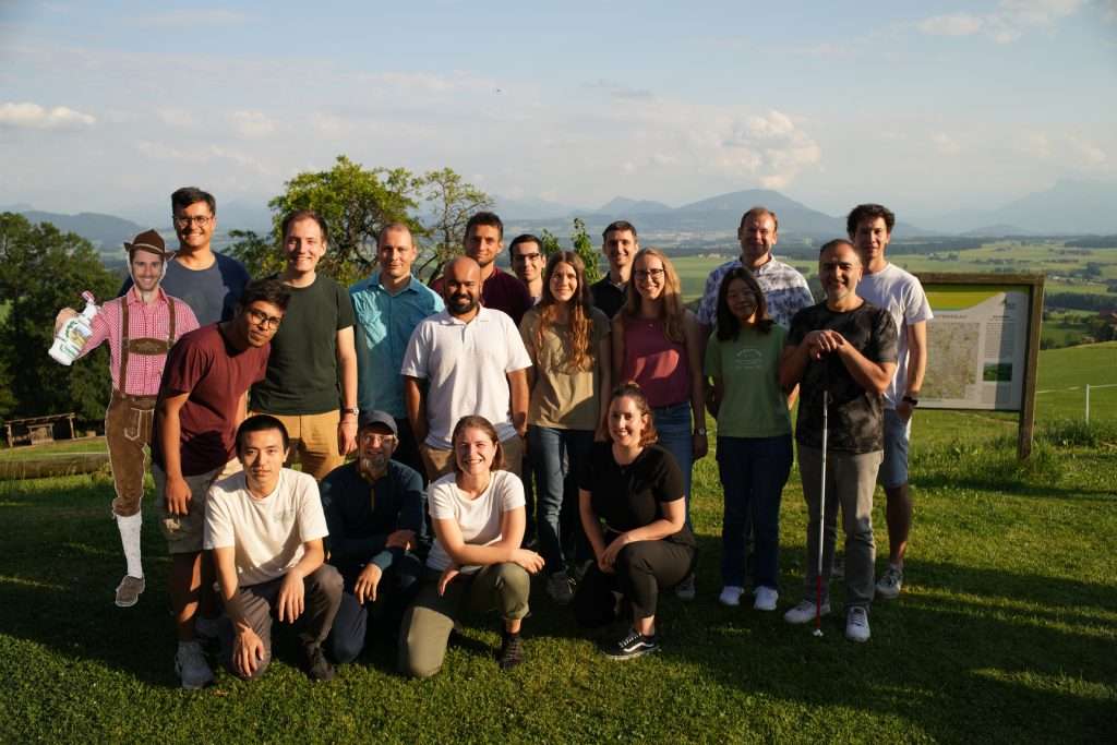
The computer vision group deals with general problems of detecting structures in images. It covers a very heterogeneous field. Among others, we research topics such as object detection, segmentation, image registration, pose estimation, image retrieval, image classification, and text recognition. Some of our current applications are document analysis, glacier segmentation, art analysis, as well as novel digitization technologies.

Current projects
Funding source: Bundesministerium für Wirtschaft und Energie (BMWE)
Project leader: ,
Project leader: ,
Prof. Dr.-Ing. Marc Aubreville
Researcher
The pathologist is still the gold standard in the diagnosis of diseases in tissue slides. Due to its human nature, the pathologist is on one side able to flexibly adapt to the high morphological and technical variability of histologic slides but of limited objectivity due to cognitive and visual traps.
In diverse project we are applying and validating currently available tools and solutions in digital pathology but are also developing new solution in automated image analysis to complement and improve the pathologist especially in areas of quantitative image analysis.
Funding source: Bundesministerium für Forschung, Technologie und Raumfahrt (BMFTR)
Project leader: ,
The goal of this project is the investigation of multimodal methods for the evaluation of interventional workflows in the operation room. This topic will be researched in an international project context with partners in Germany and in Brazil (UNISINOS in Porto Alegre). Methods will be developed to analyze the processes in an OR based on signals from body-worn sensors, cameras and other modalities like X-ray images recorded during the surgeries. For data analysis, techniques from the field of…
Project leader: , , ,
The project deals with an important aspect of art, archeology and cultural heritage: iconography and narrative
imagery. After using Computer Vision to largely recognize objects and styles, the next challenging step is to understand the semantic level of the images. The goal is the interaction between scientists and machine, not only in the form of applied science, but as a transdisciplinary exchange of methods and image theories.
Project leader: ,
Prof. Dr.-Ing. Marc Aubreville
Researcher
The goal of this project is to detect cancerous tissue in confocal lasermicroendoscopy (CLE) images of the oral cavity and the vocal cord. The current treatment of these diseases is a histological analysis of specimen and a surgical resection, which has a rather high long-term survival rate, or radiation therapy with a lower survival rate. An early detection of cancerous tissue could lead to a lowered complication rate for further treatment, as well as a better overall prognosis for patients. Further, an in-vivo diagnosis during operation could narrow down the area for the necessary surgical excision, which is especially beneficial for cancer of the vocal cords.
For this reason, we are applying methods of pattern recognition to facilitate and support diagnosis. We were able to show that these can be applied with high accuracies on CLE images.
Funding source: andere Förderorganisation
Project leader:
The analysis of the similarity of portraits is an important issue for many sciences such as art history or digital humanities, as for instance it might give hints concerning serial production processes, authenticity or temporal and contextual classification of the artworks.
In the project, first algorithms will be developed for cross-genre and multi-modal registration of portraits to overlay digitized paintings and prints as well as paintings acquired with different imaging systems such as…
Funding source: DFG-Einzelförderung / Sachbeihilfe (EIN-SBH)
Project leader: , ,
Funding source: DFG-Einzelförderung / Sachbeihilfe (EIN-SBH)
Project leader:
Dr.-Ing. Vincent Christlein
Researcher
Although OCR-D made huge progress in the last project phase in providing OCR for early printed books, it still faces two major problems: The huge variety of the material makes it extremely challenging to use generic OCR-models. Yet, selecting specific models is not possible as the sheer amount of material prevents a fully automatic workflow. This situation is further complicated by the lack of appropriate OCR training data. Current data sets consist overwhelmingly of texts in Fraktur, especially…
CV Colloquium
Upcoming Events:
Nina Aicher: Semi-Supervised Learning (MT Intro Talk)
Parisa Amanibeni: Investigation on Object Detection in Industrial Settings Centered on Extended Reality Platforms Through Generation and Utilization of Synthetic Data from CAD Models (MT Final)
Rahul Ramraje: Unsupervised Image Retrieval for Auction Catalogues (MT Final)
Mailing list management for students can be found here.


