These are the lecture notes for FAU’s YouTube Lecture “Medical Engineering“. This is a full transcript of the lecture video & matching slides. We hope, you enjoy this as much as the videos. Of course, this transcript was created with deep learning techniques largely automatically and only minor manual modifications were performed. Try it yourself! If you spot mistakes, please let us know!
Welcome back to Medical Engineering. So you’ve seen that we already looked into a couple of the imaging modalities in our first video and today we want to continue talking about more modalities. You will see that we talk about ultrasound and magnetic resonance imaging and more specialized modalities in this video. So looking forward to showing a couple of more modalities to you.
As promised here in part two we will have a look at a couple more imaging modalities and the first one that I want to show in this video is Magnetic Resonance Imaging.
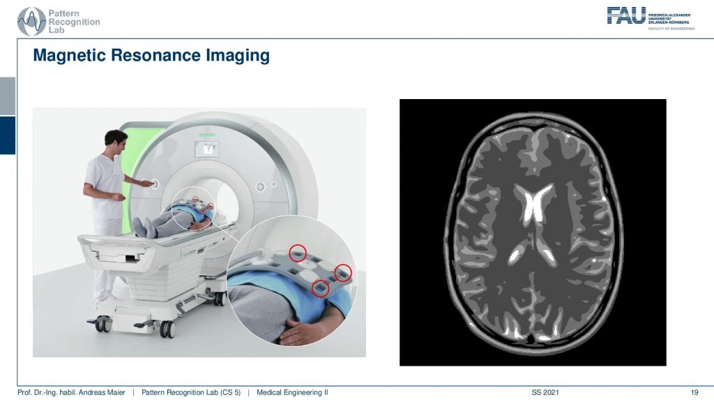
So these are scanners like the one that you see here on the left-hand side. This is a newer modality than CT. You remember computer tomography had ionizing x-rays and in this modality, we use magnetic resonance imaging. So here we are essentially using a large magnet that is applying a large external magnetic field and this magnetic field is then used in order to orient protons inside of the body into a specific direction. This then allows us to excite those protons and this is then done using radiofrequency radiation. These rf pulses are sent into the body but they are then emitted back out of sight of the body. The nice thing with rf pulses is they are not ionizing so they are not suspected to break down your DNA and you know that with x-rays there is the suspicion that this particular modality if overused may lead to the production of cancer. So MR imaging doesn’t have this problem and what’s also great about MR imaging is the wide variety of contrasts. So here we are only showing a soft-tissue contrast and you see that in the scan you can visualize the different tissues of the brain and this is much better than in CT imaging. So you can resolve the different soft tissues in the body much better. Note also that you don’t see the skull in this image. So this is actually the black structure just beneath the skin what you see on the very outside is the skin of the patient then there is no signal so black here on this image has a different physical interpretation than for example in the CT image. Then you have the different contrasts inside of the brain and you can differentiate different brain tissues but MR cannot just be used in the brain it can be used in essentially all parts of the body. There are specialized protocols where you can visualize different vessels. You can even visualize temperature and sophisticated physical effects using particular MR imaging sequences. Even the scanner is a miracle because you need a very strong magnetic field and this is created by an electromagnet and this one is using actually superconductors in order to generate this high power magnetic field. This means that the coils that are actually generating the magnetic field they’re embedded in for example liquid nitrogen. So what you see here in the donut shape oh it looks essentially like a CT scanner if you look at the scanner in more detail you will see that it’s much longer. So it’s much heavier than a CT scanner it has approximately a length of two meters in order to get a homogeneous magnetic field inside of the scanner. Then there are different coils on top that send out the rf pulses in order to create the image sequence. So it’s a really cool technology. You will see that we will actually need two lectures to talk about the different concepts in MR imaging and why it’s such a cool modality.
Molecular imaging is a modality that is using scanners like the one that you’re here seeing on the left hand side below.
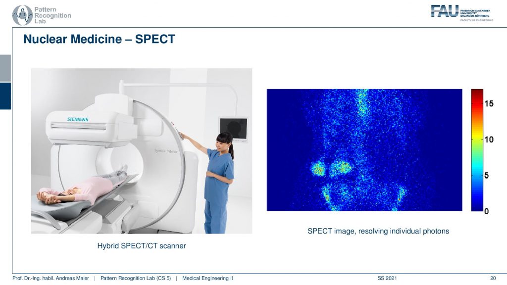
The idea is to use tracers that allow you to visualize metabolism so the function of the body. So you design specific molecules that are relevant for the metabolism and you mark them with radioactive isotopes. So you design custom molecules to image a specific part of the metabolism and these radioactive isotopes they break down and because they break down they generate radiation. So what you see here on the left-hand side is a so-called SPECT scanner. So it’s a Single Photon Emission Computed Tomography scanner and this scanner is using a particular kind of decay in order to generate the signal. The nice thing with this kind of tracer technology is that you can create essentially all kinds of molecules that are relevant for the metabolism and image the concentration of that specific molecule inside of the body. So what you see here on the right-hand side is not just an image that shows you something about the structure. It actually doesn’t show you a lot of structure at all. But you’re interested in the distribution of the specific tracer molecule and this is shown here in the image on the right-hand side. Of course, you can design tracers that range from a gas type of tracers that can be used for imaging the lung but you can also image the entire metabolism by different kinds of tracers that are swallowed and they’re digested. You can even inject them. So there are all kinds of tracers that use specific carrier molecules in order to image metabolism.
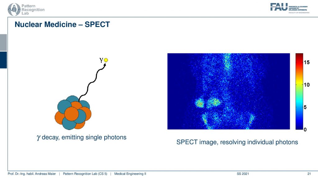
So here you see the principle of SPECT. So here you have the γ decay so this is just emitting a single photon in the radioactive decay process. You know that there is α, β, and γ decay. The γdecay here is responsible to create the image and then you have a sophisticated detector technology that then is able to create the image on the right-hand side. There’s also other decay.
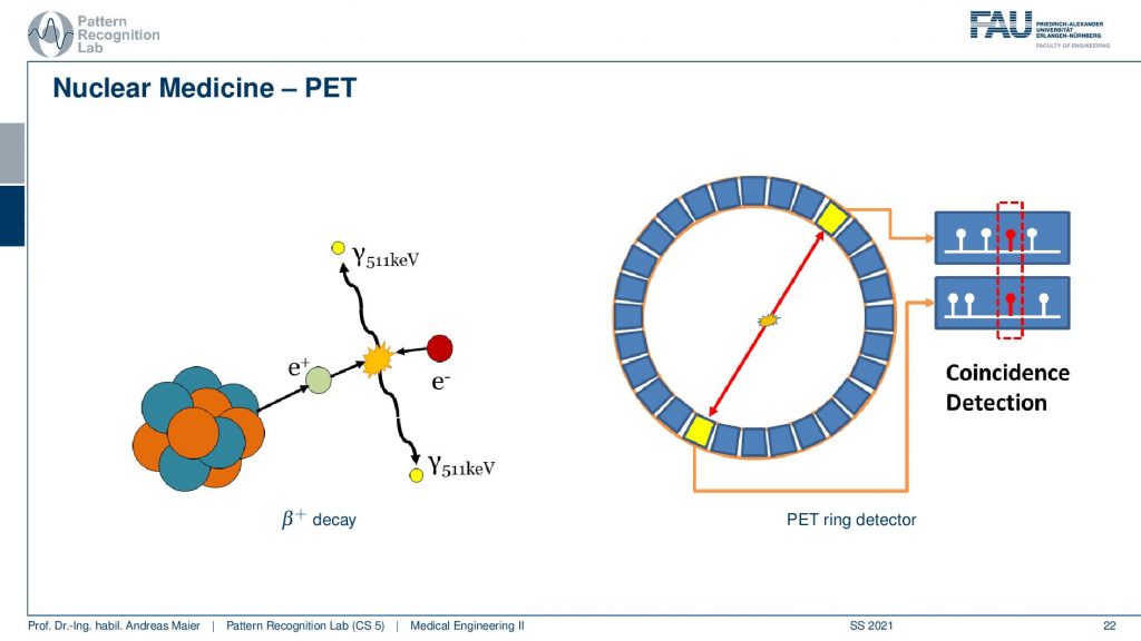
So here you see a β+ decay and in the β+ decay, you see that a positron is generated. It’s not the Positron itself that is generating the signal but the positron hits an electron they get annihilated and then two γ quanta are generated that emerge in exactly opposite positions. Because of that, you can then detect the two quanta in a ring-type of detector as you see here on the right-hand side. There is something called coincidence detection and with the coincidence detection, you can then reconstruct the ray that is connecting the two elements. So the β+ decay must have happened on this line. If you measure a sufficient number of these decays, then you’re able to reconstruct an image, and the entire technology is then called PET(the positron emission tomography).
Well, what else do we have?
There is of course ultrasound imaging. This is a quite popular imaging modality and you may have seen that for many different purposes so you have the ultrasound probe that you put on the body.
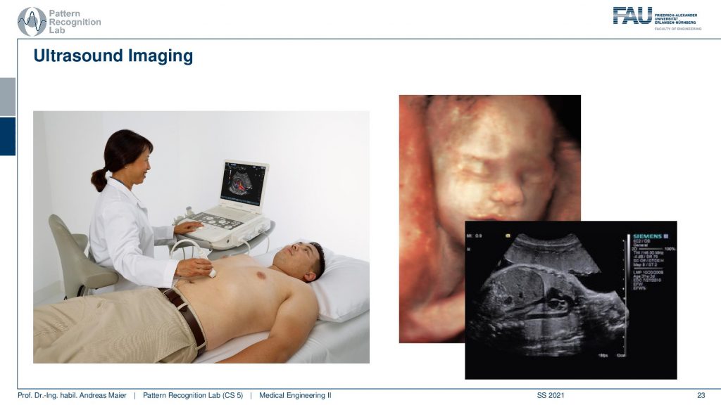
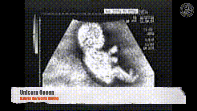
It’s very important that there’s no air between the probe and the body surface. So this is why people are using this kind of liquid gel and this gel is then used in order to prevent any air inclusions between the probe and the body and with that you can image inside of the body. It’s quite frequently used for looking at the abdominal organs and it’s also very frequently used during pregnancy where you can then image the child as it matures before birth. With 3d ultrasound technologies, you can even already reconstruct the face of the fetus such that you can get to know your future child already before birth. Ultrasound is a very popular technology and the cool thing about ultrasound is it’s not as expensive as the other modalities that we’ve seen. Probably endoscopy and ultrasound are the cheapest ones. Ultrasound is so much miniaturized by now that you can even use mobile systems that have the size of a handheld game console. So these systems are deployable you can even use them within an ambulance. So a really cool technology that can be used to make diagnoses very very quickly. So also no ionizing radiation here because we’re using as you might have guessed from the name sound signals in order to reconstruct the images.
So you see we have plenty of cool technologies coming up.
Let’s summarize them a little bit. So we’ve seen that we have these structural modalities they’re in 2d and 3d shapes.
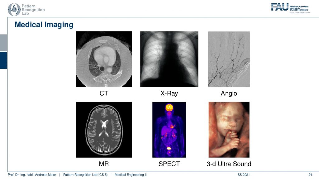
So like x-rays are 2d CT is 3d and then we also have seen that there are things like processed images like the Angiography the Digital Subtraction Angiography. We have MR images that have this sophisticated measurement set up with this very strong magnetic field. Then we have molecule-based imaging modalities like SPECT that deliver essentially the distribution of a tracer molecule and even up to things to 3d ultrasound. They are all made for different purposes. So every imaging modality is made for a different purpose and of course, people will use the best modality for that specific diagnostic purpose in order to make sure that the patient gets the best treatment possible.
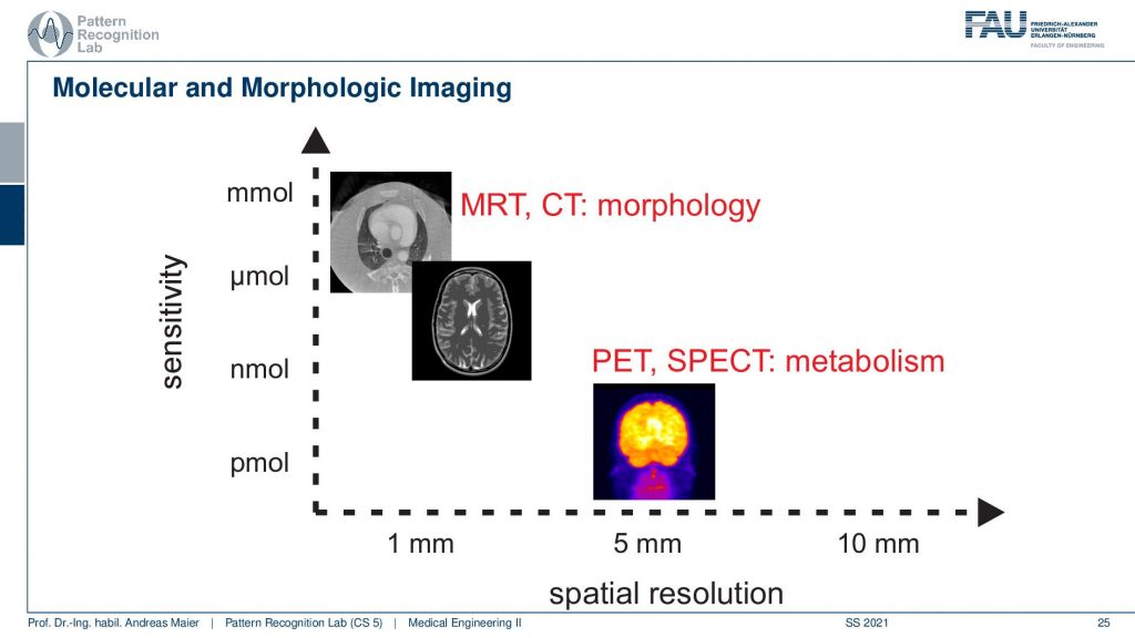
We summarize this a bit on this graph here. You can see there’s quite a bit of difference in the images and this is of course due to the image resolution and size and also the contrast that they are showing. You see that CT images that you see here on the top left have really small voxels. So you can see structures really well and they’re able to resolve really fine details. But they’re not as strong in picking up small signals. So you need a high concentration of the tissue or matter of investigation in order to be able to image contrast. So you’ve seen that you even inject contrast agent in order to get a sufficient contrast to visualize this in Angiography. Now in MR, this is a bit better as you can see the different brain tissues as we already discussed. But it comes at a cost. So the pixels are typically larger. So MR doesn’t have the same resolution as CT. So CT has more fine details and MR has the better soft-tissue resolution. This is for example when you image the brain then you may want to have a different point of optimal operation for that specific imaging modality. Then on the other side, we have the nuclear imaging modalities like here PET, SPECT. Sometimes they have really big pixels. So pixels maybe half a centimeter big. So they’re really big pixels but you can image tiny fractions of tracer. So you don’t want to inject a lot of radioactive substance into the patient right. So there’s only a couple of picomoles that actually get inserted into the body in a specific voxel and still, you are able to pick up that specific signal. So here we are operating on the far end we have a very coarse resolution, but we can see really tiny details and even specific molecules that are relevant for the metabolism for detecting cancer lesions and so on. Metastasis is typically very frequently detected if you have imaging modalities like PET and SPECT.
So you see that for every diagnostic problem-specific imaging modalities have been developed. This is essentially the idea of this class that we want to get into the different modalities, into the different diagnostic tasks. Therefore we have to talk a bit about theory. So what we will talk about in the next video is gonna be an introduction to signal processing. We will look a bit into systems theory and try to understand what the common things of all those imaging modalities are. You will see that all of them suffer the same problems from sampling from digitization, how to get into the digital domain in order to process the images then. This is why we need systems theory and we will introduce that in the next couple of videos.
I do have some further readings for you and of course, we now have the textbook which I think is pretty good. You can download it for free and then of course there’s also the book by Oppelt which is also a pretty good read. So I definitely recommend to have a look at that book as well.
This brings us to the end of this video. The next video will be about an introduction to systems theory. We will learn about concepts like convolution and the Fourier transform and you will see that they are really fundamental to signal processing and these concepts will pop up again and again throughout the entire class. This is why we look a bit into the math and the theory behind that. So I hope you enjoyed this little video and please give us feedback if you see something that we could improve on. I’m very much looking forward to seeing you in the next video bye-bye!!
If you liked this post, you can find more essays here, more educational material on Machine Learning here, or have a look at our Deep Learning Lecture. I would also appreciate a follow on YouTube, Twitter, Facebook, or LinkedIn in case you want to be informed about more essays, videos, and research in the future. This article is released under the Creative Commons 4.0 Attribution License and can be reprinted and modified if referenced. If you are interested in generating transcripts from video lectures try AutoBlog
References
- A. Maier, “Introduction”, in Medical imaging systems: an introductory guide, edited by A. Maier, et al. (Springer International Publishing, Cham, 2018), pp. 7–12, 10.1007/978-3-319-96520-8_1
- A. Oppelt, Imaging systems for medical diagnostics: fundamentals, technical solutions and applications for systems applying ionizing radiation, nuclear magnetic resonance and ultrasound, (Publicis, 2006)
Video References
- Randy Dickerson – Syngo Via VB20 Cinematic Rendering https://youtu.be/I12PMiX5h3E
- Unicorn Queen – Baby in the Womb Driving https://youtu.be/8j8iXYRWfyg