Thesis Description
Pulmonary embolism (PE) is the third most common case of cardiovascular disease, after myocardial infection and stroke [1]. According to the report of the American Heart Association in 2020, there were 370,000 cases of PE in the US in 2016 [2]. PE most commonly originate from a deep venous thrombosis in the legs, which breaks free and migrates towards the heart and lodges in the pulmonary arteries [3]. The 30-day mortality rate for PE in the US was 9.1% in 2020 [2]. Hereby, the majority of cases of preventable deaths occur due to missed diagnosis, but not due to failure of therapy [4]. Therefore, a rapid and correct diagnosis can reduce the fatality rate and is crucial for the patient’s prognosis [5, 6].
In the context of diagnostic tools for PE, computed tomographic pulmonary angiography (CTPA) has become the gold standard imaging test, as the pulmonary filling defect can be visualized with the help of contrast agent [5]. However, CTPA does not provide functional information on lung perfusion and clots in more distal branches of the lung arteries are hard to detect [7]. A rather new method, dual energy CT (DECT), counteracts these problems, as it is possible to visualize the iodine distribution in the lung parenchyma with the help of iodine maps for functional assessment of lung perfusion [8]. The use of a dual-source CT scanner allows for simultaneous image acquisition at two different energy enabling a decomposition into iodine and non-iodine, which is not possible in plain CTPA [7, 9]. The decomposition helps to understand hemodynamics in the parenchyma of interest, here the pulmonary vascular tree. However, there are drawbacks of DECT scans for PE diagnosis. A common reason for misdiagnosis of PE is the occurrence of heterogeneous perfusion due to beam-hardening artifacts from high concentration of contrast agent [7, 9]. Another problem is the accessibility of special scanners with two X-ray sources, that can execute the DECT.
In general, the performance of a CT scan with contrast agent is a time-consuming and cumbersome procedure that might require special scanners, which may not be available in every part of the world. Additionally, patients are asked to hold their breath during scan time, which can be infeasible considering children or patients that are not fully conscious [10, 11]. A non-contrast CT scan (NCCT) can be performed within seconds and is a less difficult procedure. The detection of PE in a NCCT scan is difficult but possible. Several studies showed that especially central PE could be identified in unenhanced CT scans [12, 13]. Yet, the evaluation of NCCT scans by radiologists is neither sensitive nor specific enough to reliably detect PE [14].
To guide and speed up the physicians diagnosis of PE, machine learning algorithms have been used to automatically detect PE in current research [15, 16]. These methods were developed with the intent to identify clinically important PE and prioritize worklists, considering the increasing number of e.g. CTPA scans [15]. Weikert et al. evaluated the performance of a two-staged AI-powered algorithm detecting PE from CTPA scans [15]. The algorithm includes a 3D deep convolutional neural network based on a ResNet architecture. Evaluation of the algorithm with the institution’s test set (n=1499) resulted in a sensitivity of 92.7% and specificity of 95.5% for detecting PE. Another model, PENet, was developed by Hunag et al., a 77-layer 3D convolutional neural network (CNN), evaluated on data from 2 different institutions [16]. Detecting PE, it achieved an AUROC of 0.84 on the internal test set and 0.85 on the external dataset, using the entire volumetric CTPA imaging data.
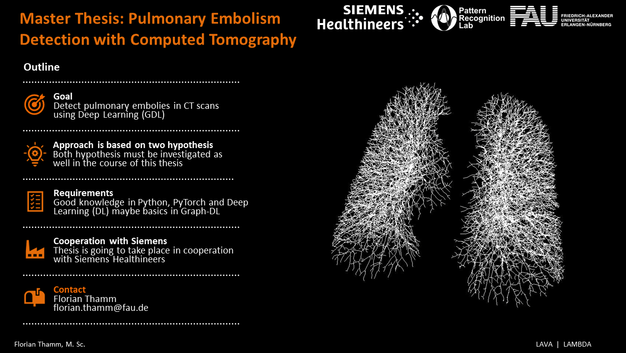
Under the aspect of availability and feasibility of contrast-enhanced CT scans, the automatic detection of PE using a NCCT scan is desirable. Additionally, modern deep learning methods could possibly exceed human performance in the detection of PE in unenhanced CT scans. Other work has already proven the success of AIbased detection of PE in CTPA data [17], whereas it is now to be investigated if the automated detection based on unenhanced CT scans is possible. Therefore, the main goal of this work is to detect PE in an NCCT scan. This thesis explores a recently published method for medical object detection, nnDetection, which is supposed to adapt itself to arbitrary detection problems [18]. Furthermore, depending on its performance on the given task, this method is extended, or other deep learning architectures are exploited alongside nnDetection. These consider the topology of the lung tree, in order to tackle the goal of PE detection in NCCT images. In addition, this work includes a detailed analysis of the models’ performances accomplishing the task of PE detection. In general, the thesis covers the following topics:
- Systematic literature research
- Detection of PE with Deep Learning
- nnDetection as baseline
- Extension of the baseline, by further improvements using prior knowledge of data and/or
- Development of other suitable Deep Learning methods
- Analysis of the Deep Learning model
- Evaluation of the results
- Comparison with baseline model
References
[1] Meredith Turetz, Andrew T. Sideris, Oren A. Friedman, Nidhi Triphathi, and James M. Horowitz. Epidemiology, Pathophysiology, and Natural History of Pulmonary Embolism. Seminars in Interventional
Radiology, 35(2):92–98, 2018.
[2] Salim S. Virani, Alvaro Alonso, Emelia J. Benjamin, Marcio S. Bittencourt, Clifton W. Callaway, April P. Carson, Alanna M. Chamberlain, Alexander R. Chang, Susan Cheng, Francesca N. Delling, Luc Djousse,
and et al. Heart disease and stroke statistics – 2020 update: A report from the american heart association. Circulation, 141(9):e139–e596, 2020.
[3] S. Takach Lapner and C. Kearon. Diagnosis and management of pulmonary embolism. BMJ (Online), 346(7896):1–9, 2013.
[4] Peter F. Fedullo and Victor F. Tapson. The Evaluation of Suspected Pulmonary Embolism. New England Journal of Medicine, 349(13):1247–1256, 2003.
[5] Waleed Abdellatif, Mahmoud Ahmed Ebada, Souad Alkanj, Ahmed Negida, Nicolas Murray, Faisal Khosa, and Savvas Nicolaou. Diagnostic Accuracy of Dual-Energy CT in Detection of Acute Pulmonary Embolism: A Systematic Review and Meta-Analysis. Canadian Association of Radiologists Journal, 72(2):285–292, 2021.
[6] Cecilia Becattini, Maria Cristina Vedovati, and Giancarlo Agnelli. Diagnosis and prognosis of acute pulmonary embolism: Focus on serum troponins. Expert Review of Molecular Diagnostics, 8(3):339–349,
2008.
[7] Long Jiang Zhang, Chang Sheng Zhou, U. Joseph Schoepf, Hui Xue Sheng, Sheng Yong Wu, Aleksander W. Krazinski, Justin R. Silverman, Felix G. Meinel, Yan E. Zhao, Zong Jun Zhang, and Guang Ming Lu. Dualenergy CT lung ventilation/perfusion imaging for diagnosing pulmonary embolism. European Radiology, 23(10):2666–2675, 2013.
[8] Dong Jin Im, Jin Hur, Kyung Hwa Han, Hye Jeong Lee, Young Jin Kim, Woocheol Kwon, and Byoung Wook Choi. Acute pulmonary embolism: Retrospective cohort study of the predictive value of perfusion defect volume measured with dual-energy CT. American Journal of Roentgenology, 209(5):1015–1022, 2017.
[9] Guang Ming Lu, S. Y. Wu, B. M. Yeh, and L. J. Zhang. Dual-energy computed tomography in pulmonary embolism. British Journal of Radiology, 83(992):707–718, 2010.
[10] Marc Rodger and Philip S. Wells. Diagnosis of pulmonary embolism. Thrombosis Research, 103(6):225–238, 2001.
[11] Konstantin Nikolaou, Sven Thieme, Wieland Sommer, Thorsten Johnson, and Maximilian F. Reiser. Diagnosing pulmonary embolism: New computed tomography applications. Journal of Thoracic Imaging,
25(2):151–160, 2010.
[12] Christopher Thom and Nathan Lewis. Never say never: Identification of acute pulmonary embolism on non-contrast computed tomography imaging. American Journal of Emergency Medicine, 35(10):1584.e1– 1584.e3, 2017.
[13] Rocco Cobelli, Maurizio Zompatori, Giovanni De Luca, Gianfranco Chiari, Paolo Bresciani, and Carla Marcato. Clinical usefulness of computed tomography study without contrast injection in the evaluation
of acute pulmonary embolism. Journal of Computer Assisted Tomography, 29(1):6–12, 2005.
[14] Simon Sun, Alexandre Semionov, Xuanqian Xie, John Kosiuk, and Benoˆıt Mesurolle. Detection of central pulmonary embolism on non-contrast computed tomography: A case control study. International Journal of Cardiovascular Imaging, 30(3):639–646, 2014.
[15] Thomas Weikert, David J. Winkel, Jens Bremerich, Bram Stieltjes, Victor Parmar, Alexander W. Sauter, and Gregor Sommer. Automated detection of pulmonary embolism in CT pulmonary angiograms using an AI-powered algorithm. European Radiology, 30(12):6545–6553, 2020.
[16] Shih Cheng Huang, Tanay Kothari, Imon Banerjee, Chris Chute, Robyn L. Ball, Norah Borus, Andrew Huang, Bhavik N. Patel, Pranav Rajpurkar, Jeremy Irvin, Jared Dunnmon, Joseph Bledsoe, Katie Shpanskaya, Abhay Dhaliwal, Roham Zamanian, Andrew Y. Ng, and Matthew P. Lungren. PENet—a scalable deep-learning model for automated diagnosis of pulmonary embolism using volumetric CT imaging. npj Digital Medicine, 3(1):1–9, 2020.
[17] Shelly Soffer, Eyal Klang, Orit Shimon, Yiftach Barash, Noa Cahan, Hayit Greenspana, and Eli Konen. Deep learning for pulmonary embolism detection on computed tomography pulmonary angiogram: a systematic review and meta-analysis. Scientific Reports, 11(1):1–8, 2021.
[18] Michael Baumgartner, Paul F. J¨ager, Fabian Isensee, and Klaus H. Maier-Hein. nnDetection: A Selfconfiguring Method for Medical Object Detection. Lecture Notes in Computer Science (including subseries Lecture Notes in Artificial Intelligence and Lecture Notes in Bioinformatics), 12905 LNCS:530–539, 2021.