NewsFriedrich Alexander University’s Pattern Recognition Lab Shines at International Symposium on Biomedical Imaging 2023
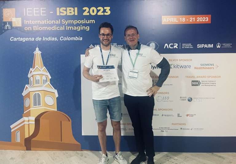
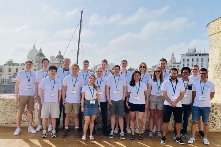
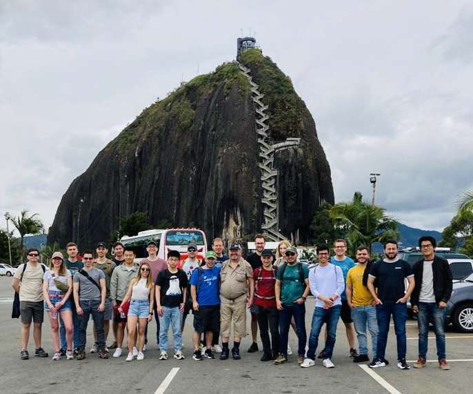
Friedrich Alexander University’s Pattern Recognition Lab has achieved great success at the 20th IEEE International Symposium on Biomedical Imaging held in Cartagena de Indias, Colombia, from April 18-21, 2023. The lab had a total of 16 papers accepted at the conference, which is a remarkable achievement as only 521 papers were accepted in total. This […]Friedrich Alexander University’s Pattern Recognition Lab has achieved great success at the 20th IEEE International Symposium on Biomedical Imaging held in Cartagena de Indias, Colombia, from April 18-21, 2023. The lab had a total of 16 papers accepted at the conference, which is a remarkable achievement as only 521 papers were accepted in total. This […]