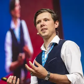
Index
From Prompt to Command: Adaptation of LLMs for Robotic Task Execution in Manufacturing
Fast heart sound detection using audio fingerprint
Style-based Handwriting Generation with LCM Diffusion Transformer
Depth-Aware Detector Localization in Freehand X-Ray Imaging
Surrogate Model for Physics Informed Lifetime Prediction in Power Electronics based on Mission Profiles
Vision-Language Models for Pathology Report Generation from Gigapixel Whole-Slide Images
Context-Aware Emotion Recognition from Pictures using Frozen CLIP
ThesisProposalVinzenzDeworDynamic Gap Closure Forecasting in the DAX Index
Evaluation and optimization of an implicit neural representation framework for markerless tumor tracking during radiotherapy
In the radiotherapy of tumors, a precise definition of the tumor volume is essential, in order to keep the radiation exposure of the surrounding tissue as low as possible. For this purpose, a planning CT scan is taken before treatment, which is used to define the area to be irradiated, also known as the planning target volume (PTV). The PTV is always chosen to be larger than the actual volume of the tumor in order to ensure that a sufficiently high dose is applied despite uncertainties such as positioning or movement of the tumor volume due to respiration. [1] Especially in the thorax and abdomen, the intrafractional movement due to respiration and physiological changes is very high. In order to compensate for this, the respiratory movement can be measured using external surrogates and imaging techniques and its extent can also be restricted using special breathing techniques. However, these methods only allow an indirect conclusion to be drawn about the tumor position. Although it is possible to measure the movement of the tumor using implanted markers, this is an additional invasive procedure, which is associated with corresponding risks and delays the start of treatment. [2] For the most accurate description of tumor movement, it would be advantageous to automatically segment the tumor on the fluoroscopic x-ray images of the linear accelerator and track its position in real-time. Since the low soft tissue contrast in the fluoroscopy projection images impedes distinguishing the tumor from surrounding structures, tracking the tumor in a synthesized 3D scan volume and then projecting its location onto the 2D x-ray image could improve the segmentation quality. Shao et al. [3] have recently presented this kind of approach with the dynamic reconstruction and motion estimation (DREME) framework. During training they divide the 3D tracking into 2 separate tasks, first a motion estimation consisting of a CNN encoder and a B-spline- based interpolant and second the reconstruction of a reference CBCT scan from the pre-treatment dynamic CBCT projections using implicit neural representations (INR). During inference, the network gets the x-ray projections as input to estimate the motion and deform the reference CBCT volume to synthesize the current real-time CBCT. [3] The goal of this thesis is to re-implement the DREME framework and evaluate its performance on our own dataset of abdominal tumors, since in their paper, Shao et al. [3] report results only on a digital phantom and a lung dataset. Furthermore their reported training time of 4 hours is not feasible in the current clinical workflow, therefore this thesis aims to explore optimization techniques, e.g. pre-training on the planning CT, to reduce the training time.
The thesis will include the following points:
- Literature review on INR deep learning methods;
- Implementation of the DREME framework for real-time motion tracking;
- Performance evaluation on our fluoroscopy dataset;
- Exploration of different strategies to reduce the training time (e.g. pre-training on the planning CT or other patients, higher parallelization, selective loss computation, etc.).
If you are interested in the project, please send your request to: pluvio.stephan@uk-erlangen.de
Prior experience in Python, Deep Learning and PyTorch is required.
Evaluation of SHViT for volumetric Semantic Segmentation in Industrial CT Scans
Industrial computed tomography (iCT) is a widely applied tool in non-destructive testing, material analysis, quality control, and metrology. Semantic segmentation of industrial CT data plays a central role in these applications by enabling quality inspection, material differentiation and part separation [1]. While convolutional neural networks (CNNs) have traditionally performed well in segmentation tasks by capturing local structures, their limited ability to model long-range dependencies poses challenges in complex 3D datasets.
Transformer-based models have recently emerged as promising alternatives. By dividing the input into patches and using self-attention mechanisms, transformers can model global dependencies. However, early vision transformers had difficulties capturing spatial structure and learning from limited data. The Swin Transformer was one of the first models to address these issues by introducing a hierarchical structure and shifted windows, combined with an inductive bias that improves generalization on small datasets [2].
Despite these advances, transformers remain resource intensive. New models such as the Shifted-window Hierarchical Vision Transformer (SHViT) aim to reduce computational costs while maintaining performance. SHViT extends the Swin architecture and improves spatial modeling and efficiency through a refined hierarchical structure with shifted windows [3].
This thesis focuses on the implementation and evaluation of a volumetric SHViT model for 3D semantic segmentation. The model is tested on a real-world dataset of industrial CT scans of boxed shoes, which includes several segmentation tasks: separating the shoes from their surroundings and identifying individual components such as the insole, outsole, and upper [4]. Typically for industrial CT data, the dataset is limited in size. Yet, its structural variability makes it an interesting benchmark for assessing model generalization. As evaluation metric for the class imbalanced segmentation dataset primarily the F1-score is used. The network is also evaluated in terms of memory and computational resource use.
The SHViT model will be compared to a CNN-based baseline, evaluating accuracy, robustness, and computational efficiency in the context of 3D industrial segmentation. While the study aims to inform the selection of neural architectures for iCT applications, its conclusions are limited using a single dataset. Nonetheless, SHViT shows potential for broader use in iCT, as it could enable the efficient application of transformer-based models to volumetric segmentation across diverse industrial datasets.
Literature
| [1] | S. a. G. P. a. V. P. a. D. W. Bellens, „Machine learning in industrial X-ray computed tomography–a review,“ CIRP Journal of Manufacturing Science and Technology, pp. 324–341, 2024. |
| [2] | Z. a. L. Y. a. C. Y. a. H. H. a. W. Y. a. Z. Z. a. L. S. a. G. B. Liu, „Swin Transformer: Hierarchical Vision Transformer using Shifted Windows,“ in Proceedings of the IEEE/CVF International Conference on Computer Vision (ICCV), Montreal, CA, 2021. |
| [3] | S. a. R. Y. Yun, „SHViT: Single-Head Vision Transformer with Memory Efficient Macro Design,“ Proceedings of the IEEE/CVF Conference on Computer Vision and Pattern Recognition (CVPR), pp. 5756–5767, 2024. |
| [4] | M. Leipert, G. Herl, J. Stebani, S. Zabler und A. Maier, „Three Step Volumetric Segmentation for Automated Shoe Fitting,“ e-Journal of Nondestructive Testing, Bd. 28, Nr. 3, 2023. |