Radiation therapy, specifically brachytherapy, is one of the most important local cancer treatment modalities for prostate cancer, enabling the delivery of a spatially varying dose distribution within a patient’s body with very high precision [1]. Brachytherapy treatment planning aims to identify the optimal treatment plan that maximally spares surrounding normal tissue structures while delivering a high dose to the target volume. For this process, a sufficient delineation of target volume, organs at risk, and identification of implanted brachytherapy needles in ultra-sound images is crucial. Deep Learning segmentation for brachytherapy treatment planning in ultrasound images is still a challenging problem, that could automate and smoothen the treatment planning process, improve standardization and reduce the treatment planning time to enable novel and improved therapies like adaptive brachytherapy [2]. Auto-segmentation of brachytherapy ultra-sound images is challenging, as ultra-sound images are noisy, have high slice-thickness and contain artifacts (e.g. from implanted needles). Based on deep learning auto-segmentations, dose calculation and optimization algorithms can be used to automatically create deliverable treatment plans [3]. The aim of this master thesis project is to create a deep learning auto-segmentation pipeline for brachytherapy ultrasound images to automate treatment planning on a cohort of more than 600 prostate cancer datasets from the Department of Radiation Oncology, University Hospital Erlangen.
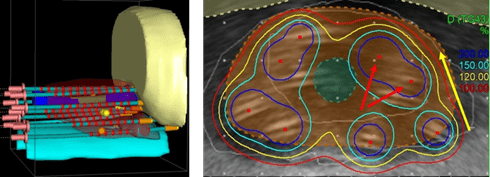
Figure 1: Overview of target volume and organ segmentation as well as needle reconstruction as performed during clinical routine (left). Structure delineation on acquired ultrasound images enables planning of the treatment dose distribution (right). Image from Karius et al. [4].
The thesis will include the following points:
- Literature review on deep learning techniques for target volume and organs-at-risk segmentation, as well needle detection on prostate Literature review on novel techniques to improve auto-segmentation perfor- mance on noisy ultra-sound images with artifacts including cardiac and fetal ultra-sound that could be trans- ferred to brachytherapy ultrasound auto-segmentation with a particular focus on integrating prior knowledge on structure shape.
- Data preprocessing for the private ultrasound treatment plan dataset including conversion of DICOM (CT, RT STRUCT) files into label maps. Automatic cleaning and splitting of the datasets into subgroups according to clinical treatment concepts using custom Python scripts (Pydicom library).
- Conversion of available needle point coordinates into binary segmentations by mapping of the known needle shape to the point coordinates.
- Training, Inference, and Testing of an nnU-net 2D and 3D fullres model on the cohort of 600 prostate can- cer ultrasound datasets to establish a baseline for comparison. Comparison of the ultrasound segmentation performance for the 2D and 3D model variant.
- Improve segmentation performance by integrating shape priors into ultrasound segmentation networks. Inte- gration of the novel method of shape prior modules (SPMs [5]) into a Unet-based segmentation Com- parative evaluation of SPM with the conventional U-net model.
- Fallback alternative / optional: Evaluate a Vision Transformer (e.g. Nnformer [6]) or a CNN-Transformer- Hybrid for brachytherapy ultrasound auto-segmentation, which could provide improved ultrasound segmenta- tion performance by improved modeling of global image context.
- Establish a pipeline for processing clinical brachytherapy ultra-sound images in DICOM format, auto-segmenting the images and exporting the auto-segmentations into DICOM RTStruct format for import into routine treatment planning programs. Evaluation of the pipeline processing time.
- Detailed evaluation of the developed brachytherapy ultra-sound auto-segmentation solution in reference to clin- ical ground truths. Perform five training and inference repeats to enable statistical analysis.
If you are interested in the project, please send your request to: andre.karius@uk-erlangen.de
Having prior experience with building neural networks in Python, especially using frameworks such as PyTorch or TensorFlow, will greatly help to develop the project.