NewsSchmidt Science Foundation awards two Computational Humanities Projects
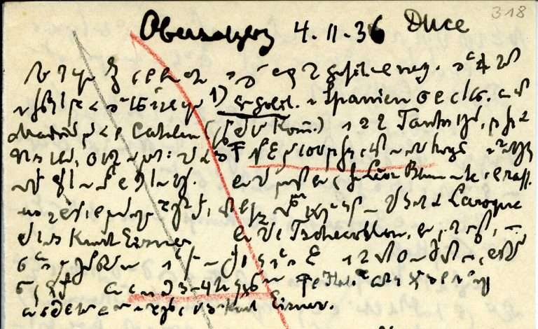
Even experts sometimes reach their limits when deciphering historical documents, for example when ancient wooden tablets are charred, scrolls have weathered, or notes are written in a rare form of shorthand. In two new projects, Vincent Christlein and his team aim to develop AI-supported technologies that make such sources easier to access. The Schmidt Sciences […]Even experts sometimes reach their limits when deciphering historical documents, for example when ancient wooden tablets are charred, scrolls have weathered, or notes are written in a rare form of shorthand. In two new projects, Vincent Christlein and his team aim to develop AI-supported technologies that make such sources easier to access. The Schmidt Sciences […]