NewsPattern Recognition Lab and EWHA Womans University Host Joint Workshop in Seoul
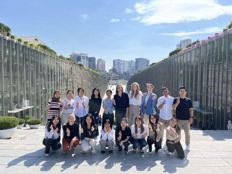
On April 22nd, the Pattern Recognition Lab and the Medical AI & Computer Vision Lab from EWHA Womans University joined forces for a workshop held on the EWHA campus in Seoul, South Korea. With a total of 10 presentations, the workshop covered a diverse array of topics spanning medical image computing, speech and language understanding, […]On April 22nd, the Pattern Recognition Lab and the Medical AI & Computer Vision Lab from EWHA Womans University joined forces for a workshop held on the EWHA campus in Seoul, South Korea. With a total of 10 presentations, the workshop covered a diverse array of topics spanning medical image computing, speech and language understanding, […]