Index

MA Intro Talk: Deep Learning for low-dose Computed Tomography CAD systems

Zeinab Meivand will hold her introduction talk in the IPA colloquium on March 7th, 12:30 p.m., in the seminar room 09.150

Supervisors:
Dr. Julian Anhaus (Siemens Healthineers, Digital & Automation Innovation)
Dr. Matthias Wolf (Siemens Healthineers, Digital & Automation Innovation)
Goldmann, Florian, M.Sc. (FAU, LME)

Thesis Description

Every year, more than two million people are diagnosed with lung cancer, demonstrating a survival rate of only 20% after 5 years after being diagnosed [1]. This leads to 1.8 million people dying from lung cancer every year, making it the leading cause of cancer-related deaths worldwide [2].

Lung cancer screening (LCS) programs have demonstrated success in decreasing the mortality when diagnosing the cancer in an early more treatable stage. Wide-scale LCS programs, such as the NLST (national lung screening trial), showed the reduction in 20% mortality in high-risk populations [3]. In direct comparison to X-ray radiography, the use of low-dose computed tomography (LDCT) has proven to be more effective for LCS, which is why upcoming nation-wide screening initiatives suggest the use of LDCT [4]. To support manual findings, experts suggest the additional use of computer-aided detection (CADe) as a secondary read to find, measure, and classify pulmonary nodules [4].

Even among experienced radiologists, there is still a moderate to high amount of inter-reader variability, depending on the size and density of individual pulmonary nodules, which affect patient management [5], but can also carry over to the both the truthing and performance of supervised learning methods. The first part of this thesis includes the assessment of the influence of the dose level (and thus the image noise) of different thoracic CT acquisitions onto the performance of different CAD systems (detection and segmentation) w.r.t the characteristics of individual pulmonary findings. Images with different dose levels will be provided. In the next step, based on the simulated dose levels, a neural network to denoise the thoracic LDCT images should be implemented. During the last years, various approaches have been introduced to apply denoising in the spatial domain, a transformed domain, and by utilizing convolutional neural networks (CNNs) and generative adversarial networks (GANs) [6] [7]. Within this work, the conventional dose level of the acquisition (ideally a full-dose scan) serves as the ground truth for the supervised learning.

Sources

[1] K. C. Thandra, A. Barsouk, K. Saginala, J. S. Aluru and A. Barsouk, “Epidemiology of lung cancer,” Contemporary Oncology, vol. 25, no. 1, pp. 45–52, 2021.
[2] H. Sung, J. Ferlay, R. L. Siegel, M. Laversanne, I. Soerjomataram, A. Jemal and F. Bray, “Global cancer statistics 2020: GLOBOCAN estimates of incidence and mortality worldwide for 36 cancers in 185 countries,” CA: a cancer journal for clinicians, vol. 71, no. 3, pp. 209–249, 2021.
[3] D. R. Aberle, A. M. Adams, C. D. Berg, W. C. Black, J. D. Clapp, R. M. Fagerstrom, I. F. Gareen, C. Gatsonis, P. M. Marcus and J. D. Sicks, “Reduced lung-cancer mortality with low-dose computed tomographic screening.,” The New England journal of medicine, vol. 365, no. 5, pp. 395–409, 2011.
[4] T. G. Blum, J. Vogel-Claussen, S. Andreas, T. T. Bauer, J. Barkhausen, V. Harth, H.-U. Kauczor, W. Pankow, K. Welcker, R. Kaaks and H. Hoffmann, “Positionspapier zur Implementierung eines nationalen organisierten Programms in Deutschland zur Früherkennung von Lungenkrebs in Risikopopulationen mittels Low-dose-CT-Screening inklusive Management von abklärungsbedürftigen Screeningbefunden,” Pneumologie, vol. 78, no. 01, pp. 15–34, 2024.
[5] S. J. Van Riel, C. I. Sánchez, A. A. Bankier, D. P. Naidich, J. Verschakelen, E. T. Scholten, P. A. de Jong, C. Jacobs, E. van Rikxoort, L. Peters-Bax, M. Snoeren, M. Prokop, B. van Ginneken and e. al., “Observer variability for classification of pulmonary nodules on low-dose CT images and its effect on nodule management,” Radiology, vol. 277, no. 3, pp. 863–871, 2015.
[6] E. Eulig, B. Ommer and M. Kachelriess, “Benchmarking deep learning-based low-dose CT image denoising algorithms.,” Medical Physics, 2024.
[7] R. T. SADIA, J. CHEN and J. ZHANG, “CT image denoising methods for image quality improvement and radiation dose reduction.,” Journal of Applied Clinical Medical Physics, vol. 25, no. 2, p. e14270, 2024.


MA intro talk Somali Roy: Generation of Artificial Vessel Trees for X-ray Image Analysis

Generation of Artificial Vessel Trees for X-ray Image Analysis

Industry advisors: Dr. Peter Fischer, Dr. Michael Manhart, Dr. Fabiola Fernández-Gutiérrez

University advisors: Prof. Dr.-Ing. habil. Andreas Maier, Florian Goldmann

Pattern Recognition Lab Members Win Third Place in AAPM’s MAR Challenge

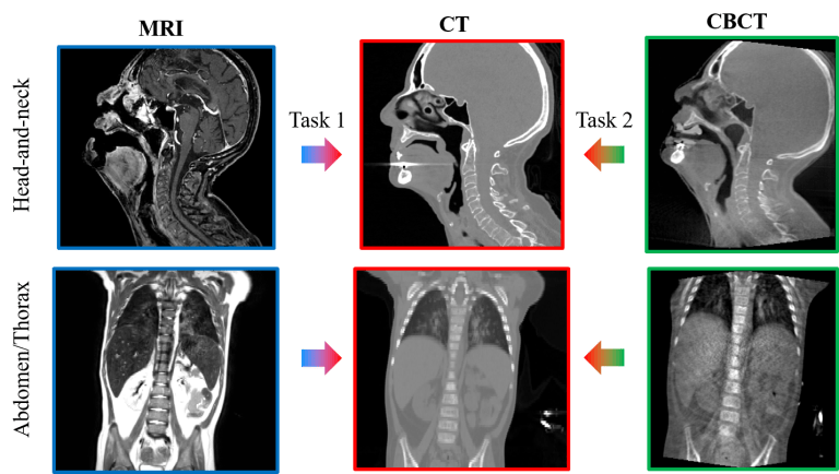
Fuxin Fan and Mareike Thies, members of the Pattern Recognition Lab, secured third place in the CT Metal Artifact Reduction (MAR) Challenge, which was organized by the American Association of Physicists in Medicine (AAPM).

The challenge focused on techniques for reducing metal artifacts in CT scans. Out of 105 registered institutions, 26 participants completed all phases of the competition. More details about the challenge can be found here: https://qtim-challenges.southcentralus.cloudapp.azure.com/competitions/1/