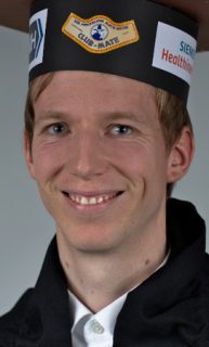
Peter Fischer
Respiratory Motion Compensation in X-Ray Fluoroscopy
Abstract
Fluoroscopy is a common imaging modality in medicine for guidance of minimally invasive interventions due to its high temporal and spatial resolution and the good visibility of interventional devices and bones. To counteract its lack of 3-D information and soft-tissue contrast, the X-ray images can be enhanced with overlays. This constitutes a medical application of augmented reality technology. Most commonly, the overlays are static. Due to inevitable respiratory and cardiac motion of the patient when imaging chest or abdomen, the images and the overlays frequently are inconsistent. In this thesis, two methods for compensating this involuntary motion are presented.
In the first method, a respiratory signal is estimated from a 2-D+t X-ray image sequence using unsupervised learning. The robustness of respiratory signal extraction to common disturbances occurring in interventional imaging is increased by patch-based processing and illumination invariance. The respiratory signal is used as prior information in the subsequent motion estimation step. Due to transparency effects in X-ray, conventional registration methods are not applicable for motion estimation. Thus, a novel surrogate-driven layered motion model is proposed to estimate the respiratory motion from the X-ray sequence. The motion model is incorporated in an energy formulation that is solved with an efficient graphics processing unit (GPU) implementation of primal-dual optimization.
In the second method, pre-operative magnetic resonance imaging (MRI) enables 3-D imaging with good soft-tissue contrast. Real-time 2-D+t slice images are acquired and stacked to 3-D+t volumes using a Markov random field (MRF). The MRF enforces similar surrogate signals in slices that are assigned to each other as well as temporal smoothness of the assignment. The surrogate signal for respiratory motion is estimated from the MRI images, while the cardiac surrogate signal is derived from electrocardiography (ECG). In the MRI volumes, conventional 3-D/3-D registration is used to estimate the patient motion. In both methods, the surrogate signals and the estimated motions are combined in a motion model, which is then used for motion compensation in X-ray.
For evaluation, experiments are conducted on pig and patient data. Compared to static overlays, the residual apparent motion in the X-ray images is reduced by 13% using the X-ray-based motion model and by 40% using the MRI-based motion model. The runtime of applying the motion model during the procedure is sufficient for real-time processing at common fluoroscopy frame rates. Two alternative methods with different properties are proposed in this thesis. X-ray-based motion compensation requires no pre-procedural processing, but is more complex during the procedure and is limited to respiratory motion. MRI-based motion compensation can also handle cardiac motion, but needs to be transferred from MRI to X-ray. The choice of motion model depends on the requirements of the clinical application. To this end, the X-ray-based motion model is implemented in a prototype and is evaluated clinically.
