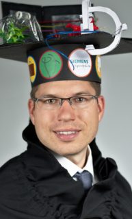
Matthias Weidler
3-D/2-D Registration of Left Atria Surface Models to Flouroscopic Images for Cardiac Ablation Procedures
Abstract
Atrial fibrillation is one of the most common cardiac arrhythmias. It can be treated minimally invasive by catheter ablation. For guidance during the intervention, augmented fluoroscopy systems gain more and more popularity. These systems allow to fuse data which was pre-operatively acquired, e.g., using computed tomography, and intra-operative patient data. This facilitates navigation during the procedure by overlaying a 3-D model of the patient’s left atrium to the fluoroscopic images. Moreover, if X-ray images are acquired from two views, 2-D image annotations can be displayed with respect to the 3-D patient model. Image fusion and annotation requires an accurate registration of pre-operative and intra-operative data which is mostly performed manually. This thesis is primarily concerned with methods for automatizing both the registration process and also steps which are required for registration. Thus, we propose also methods for reconstructing the 3-D shape of catheters from 2-D X-ray images as this is needed later for registration. In the first part of this thesis, we present methods for fast 3-D annotation of catheters. The first method is able to annotate whole line-shaped catheters in 2-D X-ray images based on a single seed point. To this end, catheter-like image regions are transformed into a graph like structure which serves as reduced search space for the catheter detection method. Resulting annotations in two X-ray images from different views can then be used to compute a 3-D reconstruction of the catheters. Our proposed method establishes point correspondences based on epipolar geometry. We define an optimality criterion that makes this approach robust with respect to spurious and missing point correspondences. Both methods are then used to establish a method for automatic cryoballoon catheter reconstruction. The second part investigates registration methods based on devices placed at certain anatomical structures. We present two different methods, one for thermal ablation and one for cryoablation. The first method relies on line-shaped devices placed outside the left atrium in the oesophagus and the coronary sinus. Their 3-D shape can be reconstructed using the algorithms presented in the first part and can then be aligned to their corresponding 3-D structures segmented from the preoperative data. The second method uses the pulmonary vein ostium which is a structure inside the left atrium in which cryoballoons are placed. A registration is established by relating the ostium position to a reconstruction of the cryoballoon computed using the approach presented in the first part. We use a skeletonization of the 3-D left atrium model to extract potential ostia from the model. In the last part we consider an automatic registration method based on injected contrast agent. In this context we present a method for classification of contrasted frames. Moreover, we present a novel similarity measure for 3-D/2-D registration that takes into account how plausible a registration is. Plausibility is determined with respect to a reconstructed contrast agent distribution within the 3-D left atrium and the contrast agent in the 2-D images. We show that a combination of this similarity measure and a measure that relates edge information from the contrast agent in 2-D images to edges of the 3-D model increases accuracy substantially. As a final step, the frame-wise registration results are postprocessed by means of a Markovchain model of the cardiac motion. This method of temporal filtering reduces outliers and improves registration quality significantly.
