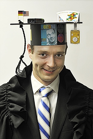
Johannes Zeintl
Optimizing Application Driven Multimodality Spatio-Temporal Emission Imaging
Abstract
Single Photon Emission Computed Tomography (SPECT) is a widely used nuclear medicine imaging technique with many applications in diagnosis and therapy. With the introduction of hybrid imaging systems, integrating a SPECT and a Computed Tomography (CT) system in one gantry, diagnostic accuracy of nuclear procedures has been improved. Current imaging protocols in clinical practice take between 15 and 45 minutes and Filtered Backprojection (FBP) is still widely used to reconstruct nuclear images. Routine clinical diagnosis is based on reconstructed image intensities which do not represent the true absolute activity concentration of the target object, due to various effects inherent to SPECT image formation.
In this thesis, we present approaches for the optimization of current clinical SPECT/CT imaging for selected applications.
We develop analysis tools for the image quality assessment of commonly used static and dynamic cardiac image quality phantoms. We use these tools for the optimization of cardiac imaging protocols with the specific goal of reducing scan time and, at the same time, maintaining diagnostic accuracy. We propose a time-optimized protocol which uses iterative image reconstruction and offers a time reduction by a factor of two, compared to conventional FBP-driven protocols. The optimized protocol shows good agreement with the conventional protocol in terms of perfusion and functional parameters when tested on a normal phantom database and in prospective clinical studies.
In addition to optimizing image acquisition, we propose a calibration method for improved image interpretation which allows to derive absolute quantitative activity concentration values based on reconstructed clinical SPECT images. In this method, we specifically take the non-stationarity of iterative reconstruction into account. In addition, we estimate the imprecision of our quantitative results caused by errors from measurement instrumentation and accumulated through the course of calibration. We could show that accurate quantification in a clinical setup is possible in phantoms and also in-vivo in patients.
We use the proposed calibration method for the quantitative assessment of dynamic processes by using time-contiguous SPECT acquisitions in combination with co-registered CT images and three-dimensional iterative reconstruction. We develop a physical dynamic phantom and establish a baseline for dual-headed SPECT systems by varying time-activity input function and rotation speed of the imaging system. We could show that, using state-of-the-art SPECT/CT systems, an accurate estimation of dynamic parameters is possible for processes with peak times of 30 seconds.
