Invited Talk (Free Registration Required) – John Boone, PhD: Three Decades of Research on Breast CT – Thu May 2nd 2024, 10:15 – 11:30 CET
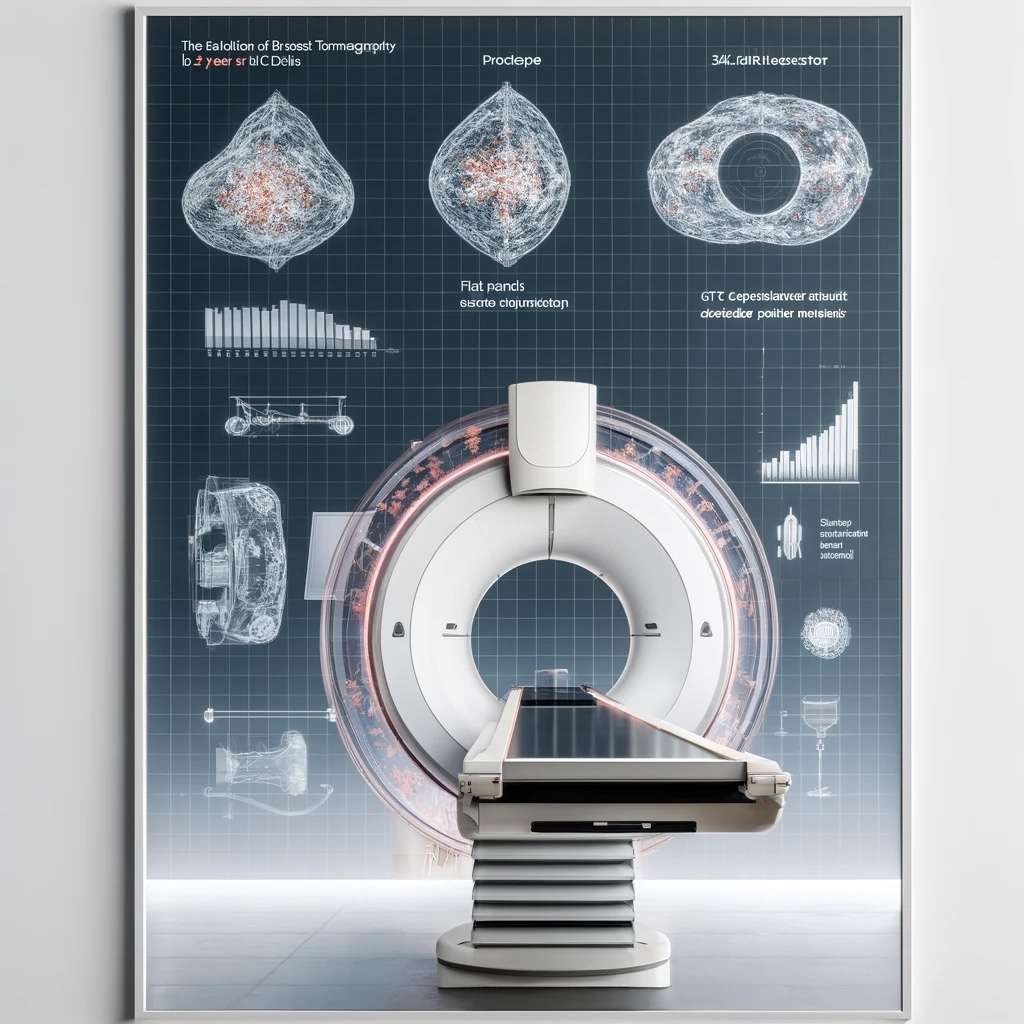
Come join us for an exciting in-person event featuring John Boone, PhD as he shares his extensive knowledge and experience in breast CT research. Discover the evolution of this technology over the past three decades, from its technical development to its current applications. Don’t miss this unique opportunity to learn from a leading expert in the field. Reserve your spot now! There will be a chance for pizza and networking right after the talk for in-person attendees.
Title: Three Decades of Research on Breast CT
Date: Thu May 2nd 2024
Time: 10:15 – 11:30 CET
Location: H14, Martensstraße 5/7, 91058 Erlangen & Online
Registration: https://www.eventbrite.de/e/invited-talk-john-boone-phd-three-decades-of-research-on-breast-ct-tickets-876654175237?aff=oddtdtcreator

Abstract: The Breast Tomography project at UC Davis began in 1999, where the feasibility of CT of the breast was established. Twenty-five years later we have designed, fabricated, and tested 4 breast CT prototype scanners in patient clinical trials. A cone beam CT system with a flat panel detector and x-ray tube is positioned under the prone patient and rotates around the patient’s breast, which is hanging pendent through a hole in the patient table. 360° of projection data (N=500) are acquired at low radiation dose, and these data are reconstructed to produce a three-dimensional volume data set with isotropic spatial resolution. Various scatter reduction algorithms have been studied, sufficient to eliminate image cupping. Monte Carlo simulations and physical measurements were used to evaluate the mean glandular dose from the procedure. Radiologist observers evaluated non-contrast and contrast-enhanced images, and more recently the terabytes of clinical breast CT image data have been used in model observer studies focused on comparing thin-slice breast imaging (breast CT) with thick-slice breast imaging (~mammography). Both synthetic mass lesions and microcalcification lesions were used with a conventional model observer (prewhitened matched filter) and AI-based (convolutional neural network) observers. These studies showed that breast CT outperforms mammography for most imaging tasks.
Short Biography: John M. Boone received his undergraduate degree in Biophysics from UC Berkeley and his doctorate degree in Radiological Sciences at University of California Irvine. After faculty positions at University of Missouri and Thomas Jefferson University, he joined the faculty at UC Davis 34 years ago. His research work has focused on the development of diagnostic x-ray imaging systems, including CT systems for specimen and breast imaging, and systems using multiple thermionic cathodes for imaging applications. He is also interested in radiation dosimetry in diagnostic imaging, and he led the effort to generate the Size Specific Dose Estimate (SSDE) metric in computed tomography, which allows more accurate dosimetry for pediatric patients and small adults. His lab developed four breast CT systems over the past 25 years and has tested their imaging performance on over 350 patients. He was board certified by the American Board of Radiology and participates in the clinical medical physics program at UC Davis. He is the co-author of the textbook “Essential Physics of Medical Imaging” with colleagues, now in its fourth edition. He currently serves as the Editor-in-Chief of Medical Physics.
Online link will be sent out one day before the talk to registered participants.
