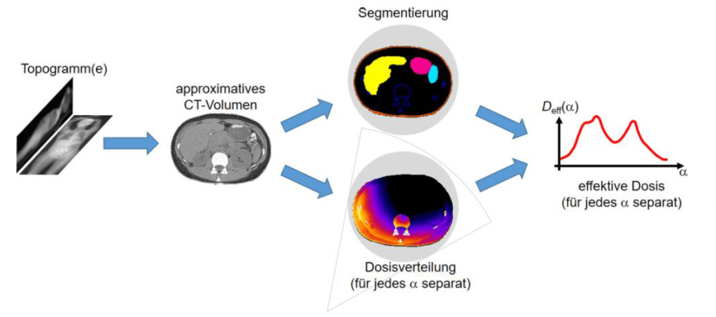
Erlangen, January 2025 – The Pattern Recognition Lab at Friedrich-Alexander-Universität Erlangen-Nürnberg (FAU) is excited to announce that the Deutsche Forschungsgemeinschaft (DFG) has granted funding for the groundbreaking project riskXSM – Advanced Dose Reduction and Spectrum Modulation in Computed Tomography (CT). The collaborative initiative has secured a total funding sum of 600,000 EUR and brings together leading experts from three institutions:
- Prof. Dr. Marc Kachelrieß (German Cancer Research Center, DKFZ Heidelberg): Real-time dose calculation and spectrum modulation.
- Prof. Dr. Michael Lell (Klinikum Nürnberg): Segmentation and patient studies using advanced simulation tools.
- Prof. Dr. Andreas Maier (FAU Erlangen-Nürnberg): Neural networks for reconstructing contrast medium distribution.
Project Goals and Vision
The primary aim of the riskXSM project is to develop a novel method for X-ray tube current and spectrum modulation in CT imaging. This approach seeks to significantly reduce radiation exposure for patients while maintaining or even enhancing image quality. This innovation promises a safer and more efficient diagnostic process for millions of CT scans performed globally each year.
The project builds on the success of its predecessor, riskTCM, which demonstrated substantial dose reduction through software-based tube current modulation. With riskXSM, the team aims to push the boundaries further by introducing spectrum modulation, an approach requiring careful optimization of tube voltage and prefilter thickness. These advancements could enable dose reductions of up to 30% or more beyond the achievements of riskTCM, making CT imaging safer and more accessible.

Technical Innovations
The project involves a combination of cutting-edge simulation techniques, phantom measurements, and advanced neural networks to predict the distribution of iodinated contrast media. These predictions are crucial for balancing radiation dose, image noise, and contrast enhancement, enabling optimal adjustments in X-ray spectrum and current. Key highlights include:
- Development of realistic simulations to validate the proposed method without requiring modifications to commercial CT systems.
- Integration of advanced neural networks to reconstruct pseudo-contrast medium distributions from limited data, leveraging expertise from prior work on synthetic contrast enhancement in the brain.
- Deployment of the ReconCT software at Klinikum Nürnberg to process patient raw data, enabling clinical evaluation of the proposed methods.
Impact on Medical Imaging
By achieving significant dose reductions or enhancing image quality without increasing radiation risk, riskXSM represents a critical step forward in patient-centered imaging. The project aligns with global efforts to minimize radiation exposure in medical diagnostics while improving diagnostic accuracy and reliability.
Prof. Dr. Andreas Maier emphasized the importance of this work:
“With riskXSM, we aim to redefine the balance between image quality and patient safety in CT imaging. This project exemplifies the power of interdisciplinary collaboration in addressing real-world challenges in medical technology.”
Acknowledgment
The Pattern Recognition Lab expresses its gratitude to the DFG for supporting this transformative research. We also thank our partners, Prof. Dr. Marc Kachelrieß and Prof. Dr. Michael Lell, for their invaluable contributions to this collaborative endeavour. Last but not least, we express our gratitude to Chang Liu whose ideas and contributions have been fundamental to the success of this project application.

